
|
Fig. S2
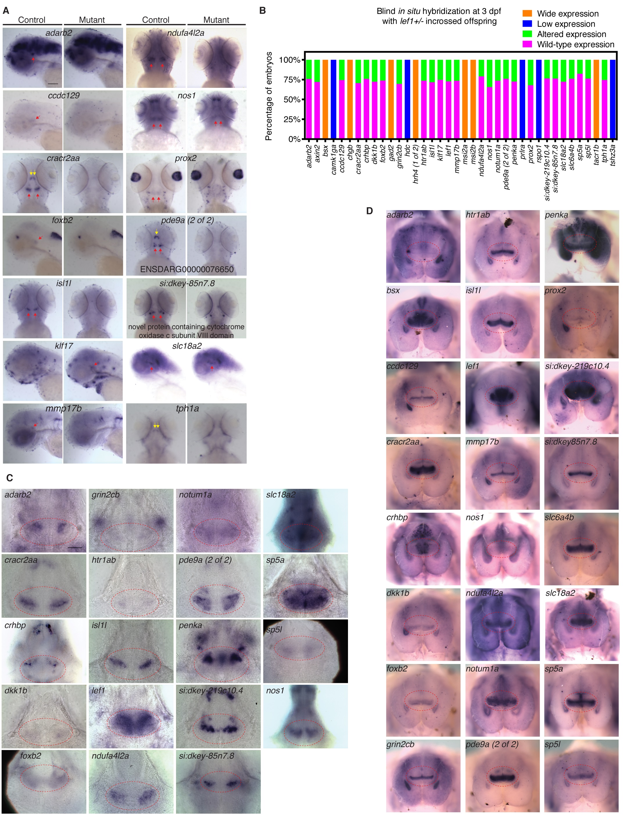
Whole mount in situ hybridization for zebrafish Lef1-dependent genes identified from RNA-seq.
(A) Representative images of whole mount in situ hybridization on 3 dpf control and lef1 mutant embryos. Red and yellow arrows indicate gene expression in caudal and rostral hypothalamus, respectively. Lateral (adarb2, ccdc129, foxb2, klf17, mmp17b, and slc18a2) or ventral (other genes) views were selected for optimal expression visualization. (B) Quantification of expression following whole mount in situ hybridization on 3 dpf offspring from lef1+/- incrosses. Fifty to eighty-five embryos were analyzed per gene. (C) Images of 3 dpf control brains centered on Hc from ventral view. (D) Gene expression in the hypothalamus of 4 months post-fertilization (mpf) female wild-type zebrafish from ventral view. Representative images are shown in (C) and (D) for at least 2 samples tested. Images of ventral view have anterior on top; images of lateral view have dorsal on top and anterior on the left. Red dashed outlines in (C) and (D) depict the caudal hypothalamus. Scale bars: 0.1 mm in (A); 5 μm in (C); 0.2 mm in (D). Raw data can be found in S1 Data.