
|
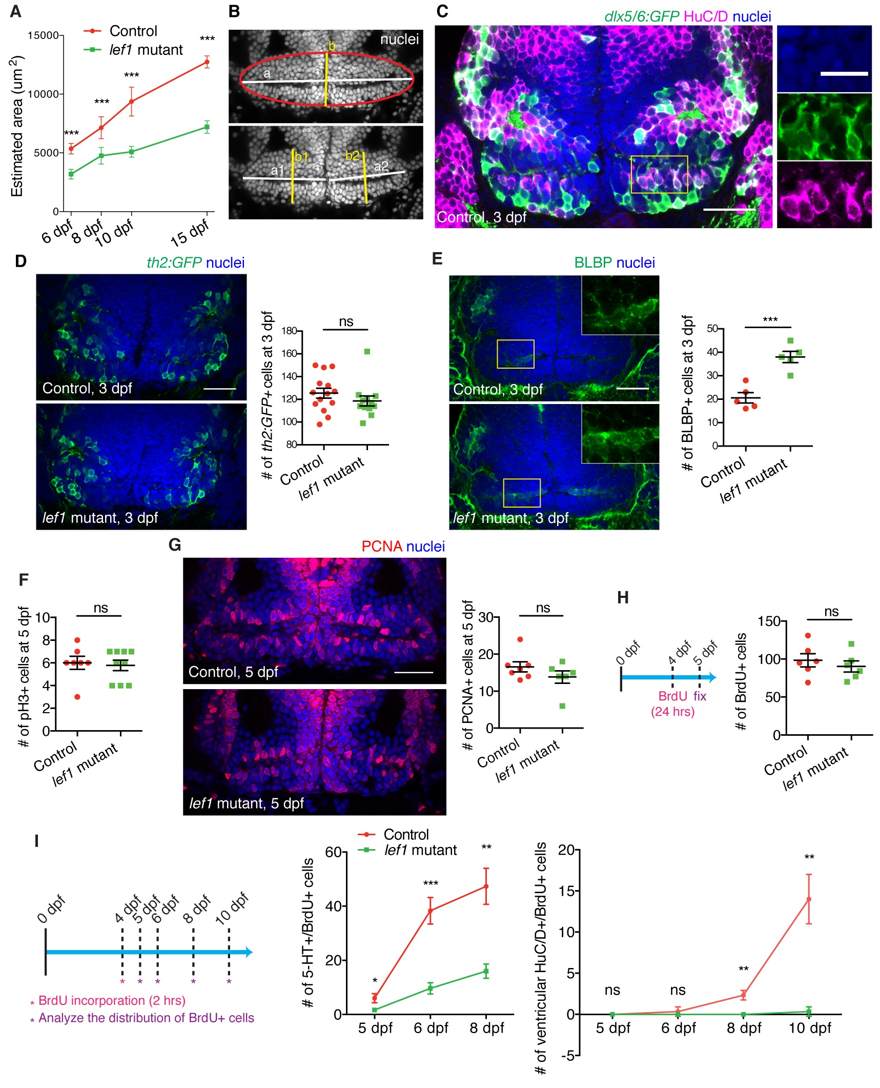
Fig. S1
Lef1 promotes neurogenesis in the zebrafish caudal hypothalamus (Hc).
(A and B) Hc size in control and lef1 mutants (A) estimated by the area of confocal ventricular slice (B). Hc was defined as an oval indicated by red outline in (B). The lengths of a1, a2, b1, b2 in the representative image (B) were measured by ImageJ, and the area of the oval was calculated by the following equations: Estimated area = π*a*b/4; a = a1+a2; b = (b1+b2)/2. (C) Co-immunostaining of HuC/D and GABAergic lineage marker dlx5/6:GFP [83] in the 3 dpf Hc. Three confocal channel-split magnified images of the region depicted by the yellow rectangle are shown on the right. A representative image is shown for at least 3 embryos tested. (D and E) Immunostaining of th2:GFP+ (D) and BLBP+ cells (E) in the Hc of 3 dpf control and lef1 mutant. Representative images are shown on the left, and quantifications are shown on the right. Higher magnification views of yellow rectangles in single channel are shown in the insets in (E). (F-H) Measurement of proliferation in the Hc of 5 dpf control and lef1 mutant as shown by pH3+ (F) and PCNA+ cells (G; representative image on the left and quantification on the right; cells adjacent to the horizontal ventricle were counted), and 1 day BrdU labeling (H; schematic on the left). (I), BrdU pulse-chase (schematic on the left) to measure birth of 5-HT+ and ventricular HuC/D+ cells after 4 dpf. Data are mean ± SEM, except mean ± SD in (A) and (I). ***P < 0.001, **P < 0.01, *P < 0.05, ns. P > 0.05 by unpaired Student t tests. All images are confocal ventricular slices. All scale bars are 25 μm except 12.5 μm in the magnified image in (C). See S1 Table for description of quantification and experimental n. Raw data can be found in S1 Data.