
|
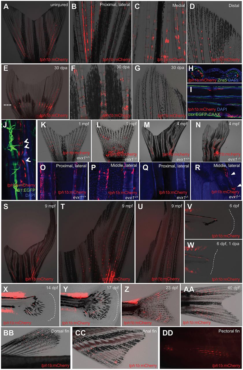
Fig. 1
tph1b+ fibroblasts localize to adult zebrafish fin joints. (A-D) tph1b:mCherry expression in uninjured adult zebrafish caudal fins. (A-D) Whole fin (A), and magnified proximal dorsal rays (B), medial rays (C) and distal mediolateral rays (D). tph1b:mCherry is expressed near fin joints. (E-G) tph1b:mCherry expression in whole (E), medial (F) or distal (G) caudal fins at 30 days post-amputation (dpa). Dashed line indicates the amputation plane. (H) Transverse section of uninjured tph1b:mCherry rays, co-stained with DsRed (red, tph1b:mCherry), ZNS-5 (osteoblasts, green) and DAPI (nuclei, blue). (I,J) Optical section through uninjured tph1b:mCherry; osx:EGFP-CAAX (I) or tph1b:mCherry; fli1:EGFP (J) fin ray, stained with DAPI (nuclei, blue). tph1b:mCherry-expressing cells do not colocalize with osteoblast markers or fli1:EGFP-expressing cells (arrowheads, J). (K-N) Wholemounted tph1b:mCherry caudal fins in wild-type (K,M) or jointless evx1−/− (L,N) background at 1 (K,L) or 4 (M,N) months post-fertilization (mpf). (O-R) Optical sections of tph1b:mCherry fin rays in a wild-type (O,P) or evx1−/− (Q,R) background, showing proximal (O,Q) or middle (P,R) lateral rays stained with DAPI (nuclei, blue). tph1b:mCherry localizes to proximal lateral rays and middle lateral rays near mature joints, but is absent in jointless fins, except for injury-induced expression (R, arrowheads). (S-U) tph1b:mCherry expression in the longfin (lof) background, shown at 9 mpf in lateral (T) and distal (U) ray tissues. (V,W) tph1b:mCherry expression at 6 dpf (V) or at 6 dpf, 1 dpa (W). Expression is not induced in larval finfold tissue after injury. Dashed lines indicate the tailfin boundary. (X-AA) tph1b:mCherry expression during caudal fin development at 14 (X), 17 (Y), 23 (Z) and 40 (AA) dpf. Dashed lines indicate fin boundary. (BB-DD) tph1b:mCherry expression in adult dorsal (BB), anal (CC) and pectoral (DD) fins, excluding distal segments.