
|
Fig. S5
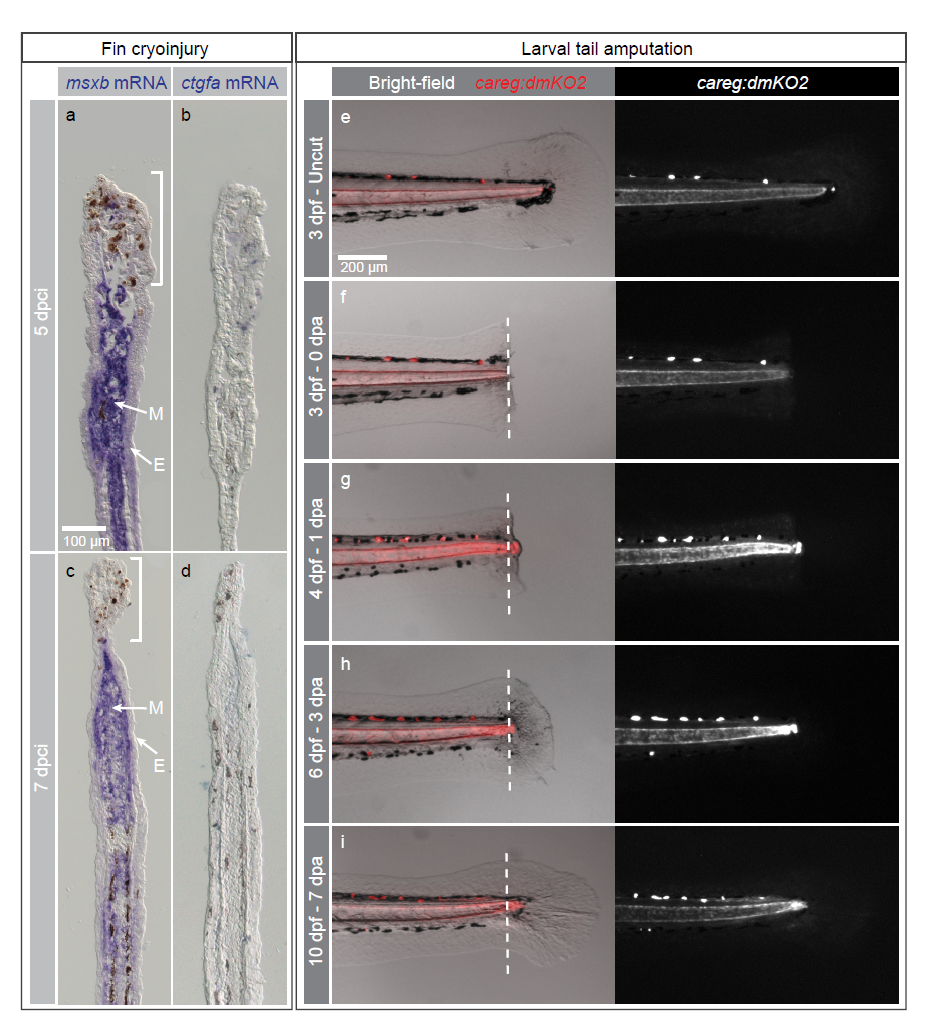
The careg reporter is activated in other injury models of tissue regeneration in zebrafish.
(a-d) In-situ hybridization (purple) on longitudinal fin sections reveals that, as in the amputation model, the endogenous ctgfa gene is not upregulated in the regenerating fin after cryoinjury. (a, c) A probe against a blastema marker, msxb, was used as a positive control to detect the activated mesenchyme. (b, d) A ctgfa probe does not label the regenerating fin tissues. M, mesenchyme; E, epidermis. Brackets indicate the damaged tissue remaining at the tip of the cryoinjured stump. N=3. (e-i) Live-imaging of the larval tail of careg:dmKO2 fish at different days post-fertilization (dpf) and post-amputation. The transgenic reporter is constitutively expressed in the notochord. After amputation (dashed line), the expression of careg:dmKO2 is markedly upregulated at the tip of the regenerating tail. N=6.