
|
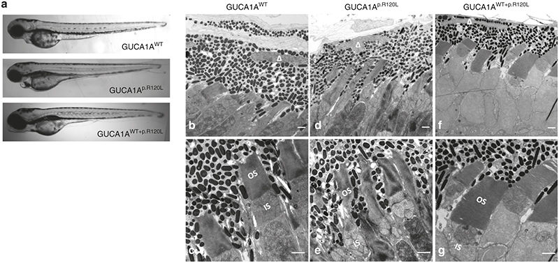
Fig. 4
Morphological changes caused by GUCA1A p.R120L. (a) Light microscopy indicates no evident morphological changes in eyes of zebrafish injected with GUCA1AWT, or GUCA1Ap.R120L, or GUCA1AWT+p.R120L 4 days postfertilization (dpf). (b–e) Transmission electron microscopy of the retina of zebrafish 11 dpf injected with GUCA1AWT or GUCA1Ap.R120L. Photoreceptors are regularly lined and shaped in the GUCA1AWT-injected group (b, d). Thickness of the RPE; shrinking, twisty, and caducous photoreceptor outer segments; and choriocapillary disruptions are found in the GUCA1Ap.R120L-injected group (c, e). ▵ represents the RPE nucleus. Scale bar = 1 μm.