
|
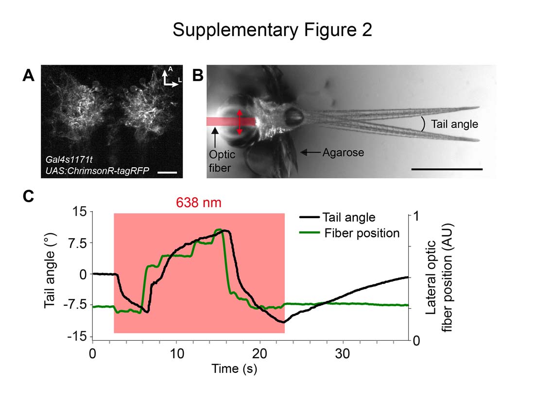
Fig. S2
Stimulation of ChrimsonR-expressing cells elicits behavior in larval zebrafish. (A) Transgenic ChrimsonR-tagRFP expression in the nucleus of the medial longitudinal fasciculus (nMLF) of a 5 dpf zebrafish larva. Scale bar, 20 μm. (B) Optogenetic setup as published previously1. The head of the same fish from (A) is embedded in agarose with its tail freed. A 50 μm optic fiber targets 638 nm light (0.1 mW) onto the region of the nMLF and is moved in a vertical direction. A projection of the initial and maximum left deflected tail positions is shown. Scale bar, 1 mm. (C) Measurement of the tail angle and the fiber position over time of the fish shown in (B). The red rectangle depicts the epoch of 638 nm light stimulation.