
|
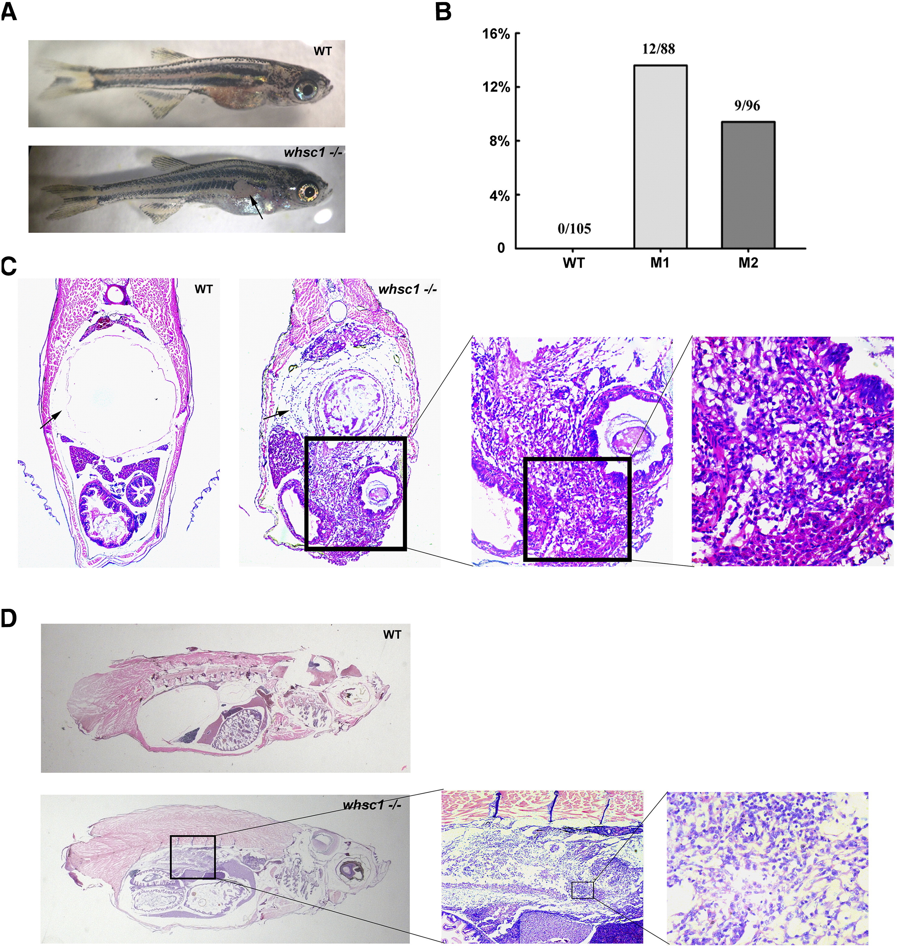
Fig. 5
whsc1 mutant zebrafish developed swim bladder lesions. (A) One-month-old whsc1−/− zebrafishes developed skin lesions. Arrow pointed to the lesion. (B) The Statistics of wild-type and whsc1 mutant fishes with lesions. (C) HE staining of swim bladder in one-month-old wild-type and whsc1 mutant zebrafishes (cross-section). Tissues were serially sectioned from head to tail, and sections containing swim bladders (wild-type fishes) or skin lesions (mutant fishes) were HE stained for analysis. (D) HE analysis of swim bladder in three-month-old zebrafish (vertical section). Note that wild-type fish had normal and functional swim bladder, while whsc1 mutant fishes had no obvious swim bladder. The position of swim bladder in whsc1 mutant was occupied with cell masses that seemed to have no cell polarity.