
|
Fig. 3
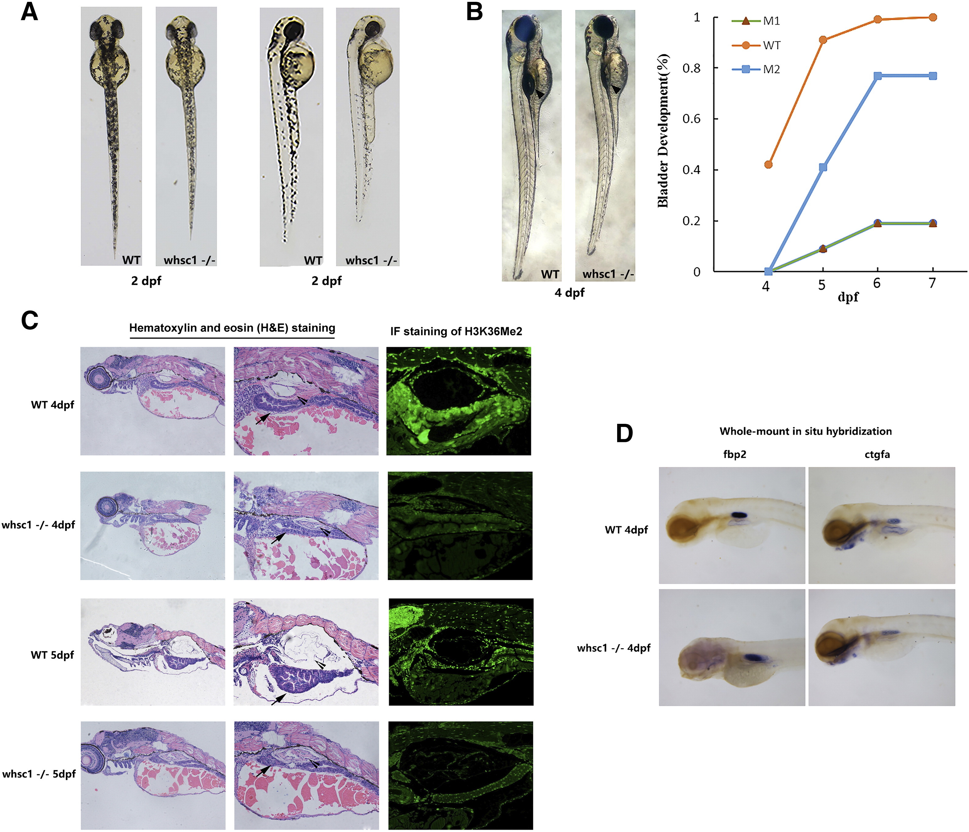
whsc1−/− zebrafishes had developmental defects in swim bladder. (A) In the early development, compared to age-matched wild-type embryos, whsc1−/− embryos showed reduced body length and less pigmentation. (B) The left part shows that swim bladder inflation defects in whsc1−/− zebrafish. Arrow heads point to the swim bladders. Note that wild-type larva had already inflation, while whsc1−/− larva had not, suggesting their swim bladders were either developed abnormally or not functional. And the right part is the statistics of inflation of swim bladder in whsc1−/− zebrafish and wild-type zebrafish. (C) HE staining and immunofluorescence analysis of the swim bladder in whsc1−/− zebrafish and wild-type zebrafish at 4 dpf and 5 dpf. Arrow heads pointed to the bladder and arrows pointed to the gut. In the HE staining of zebrafish larvae at 4 and 5 dpf (left and middle panels), bladders in wild-type larvae had formed a three layer structure that was filled with air. However, bladders in whsc1 mutants were still cell masses and failed to develop three layers. Immunofluorescence staining of the H3K36me2 revealed that whsc1−/− larvae expressed significantly lower level of H3K36me2 than wild-type larvae did. (D) WISH analysis of swim bladder markers fbp2 and ctgfa in whsc1−/− zebrafish and wild-type zebrafish at 4 dpf.