
|
Fig. S13
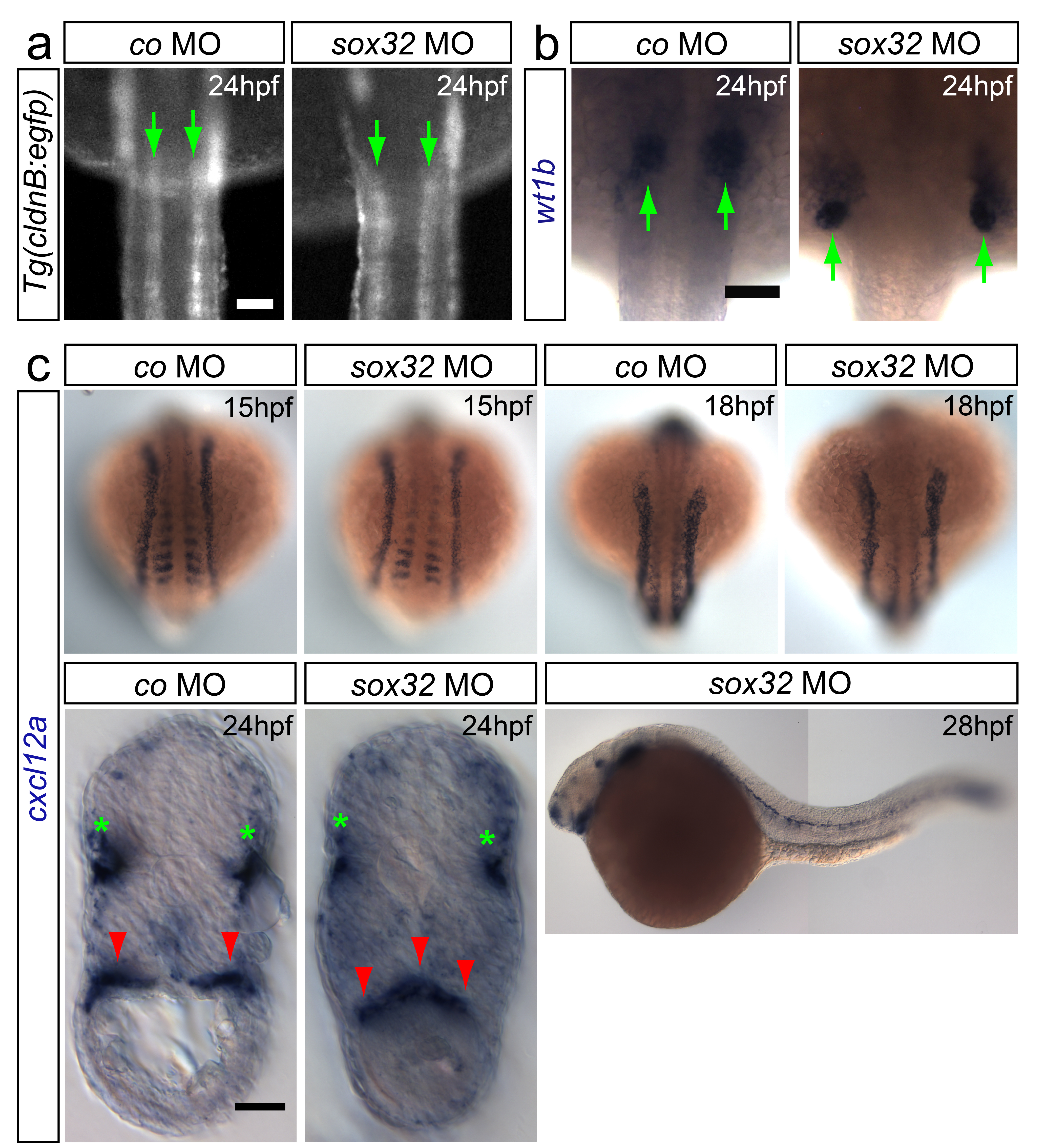
Patterning of the mesodermal tissues adjacent to the gonad is normal in gut-deficient embryos
(a-b) Bilateral arrangement of pronephric ducts and the pronephros in embryos deficient for the gut is maintained (pronephric ducts labeled using (Tg(cldnB:egfp)) and pronephros with wt1b probe, green arrows). Anterior is up. (c) Bilateral cxcl12a expression is detected in the somites and lateral plate mesoderm of both control and gut-deficient embryos at 15 and 18hpf. At 24hpf cxcl12a expression at the gonad area is altered in embryos lacking the gut (additional red arrowhead in the cross-section). Green stars indicate cxcl12a expression in the lateral line. The expression of cxcl12a at the gonad region in embryos lacking the gut tube is no longer detected at 28hpf. Scale bars in all panels 50μm.