
|
Fig. S2
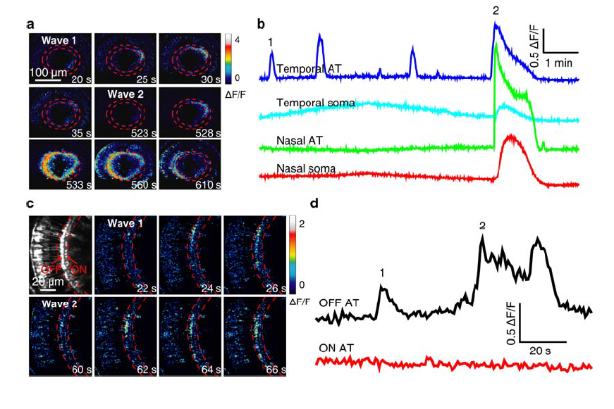
Calcium wave activities in the soma and AT of BCs.
(a) Pseudocolor time-lapse images showing two BC calcium waves (1 and 2) in a 3-dpf Tg(Gal4-VP16xfz43,UAS:GCaMP1.6) larva. The wave 1 only propagated locally at BC ATs in the temporal-dorsal retina, while the wave 2 propagated globally to the entire IPL and evaded to BC somata.
(b) Spontaneous BC calcium activities at four different retinal regions. BC ATs at the temporal retina displayed several wave-like events. The first and last waves are showed in (a).
(c) Pseudocolor time-lapse images showing two BC calcium waves (1 and 2) in a 3-dpf Tg(Gal4-VP16xfz43,UAS:GCaMP1.6) larva. Both the two waves started and propagated locally at OFF ATs in the nasal retina. Left top, fluorescence image showing OFF and ON ATs which are located at the sublaminae a and b of the IPL, respectively.
(d) Spontaneous BC calcium activities at OFF ATs, but not ON ATs in the nasal retina.
Corresponding images are showed in (c).