
Image
|
Figure Caption
Fig. S10
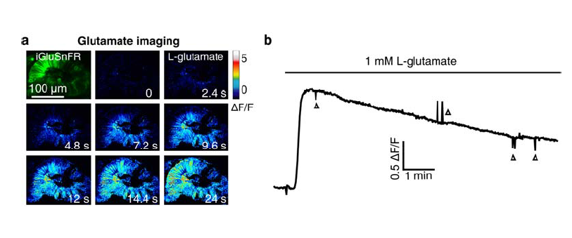
Application of L-glutamate induces an increase of glutamate signal in iGluSnFR-expressing retinal cells.
(a) Pseudocolor time-lapse images showing the change of glutamate signal before and after bath application of 1-mM L-glutamate in the retina of a 44-hpf larva expressing the glutamate biosensor iGluSnFR (Left top). The lens of the retina was removed for the diffusion of L-glutamate into the retina.
(b) Time course of glutamate signal changes in the retina (a). The downward and upward deflections (triangles) were due to the movement of the larva during imaging.
Acknowledgments
This image is the copyrighted work of the attributed author or publisher, and
ZFIN has permission only to display this image to its users.
Additional permissions should be obtained from the applicable author or publisher of the image.
Full text @ Nat. Commun.