
Image
|
Figure Caption
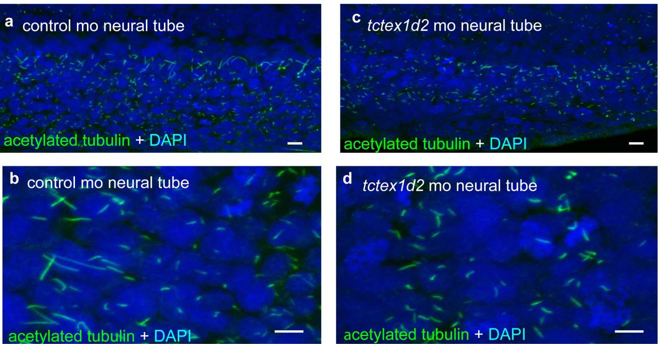
Fig. S6
Ciliogenesis appears undisturbed in tctex1d2 morphant neural tube.
The appearance of neural tube cilia in control-morpholino (a, b) and tctex1d2-morpholino (c, d) injected zebrafish embryos was similar at 24hpf using whole mount confocal microscopy after staining with anti-acetylated tubulin antibody and DAPI (see online methods for details). Scale bar 10 μm.
Figure Data
Acknowledgments
This image is the copyrighted work of the attributed author or publisher, and
ZFIN has permission only to display this image to its users.
Additional permissions should be obtained from the applicable author or publisher of the image.
Full text @ Nat. Commun.