
|
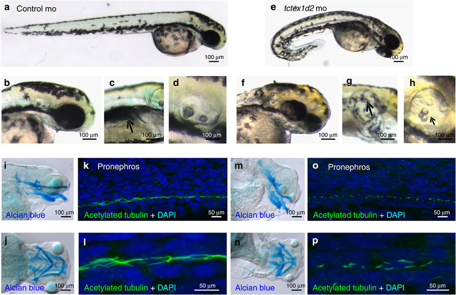
Fig. 3
Knockdown of tctex1d2 in zebrafish leads to a typical ciliopathy phenotype.
Whole-mount light microscopy showing control morpholino (mo)-injected embryos (a–d) and tctex1d2 morphants at 4 days post fertilization (e–h). Compared with controls, knockdown of tctex1d2 results in ventrally curved body axis (a,e), small eyes (b,f), pronephric cysts (c,g) and otolith defects (d,h). Alcian blue staining of cartilage identifies craniofacial cartilage defects in tctexd2 morphants (m,n) compared with controls (i,j). Immunofluorescence analysis after staining of cilia at 24 h.p.f. with anti-acetylated tubulin antibody reveals shorter cilia in the pronephric duct of tctex1d2 morphants (o, magnified in p) compared with control embryos (k, magnified in l); however, this difference was no longer evident at 48 h.p.f. (data not shown). Scale bars, 100 μm (a–j,m,n) or 50 μm (k,l,o,p).