
|
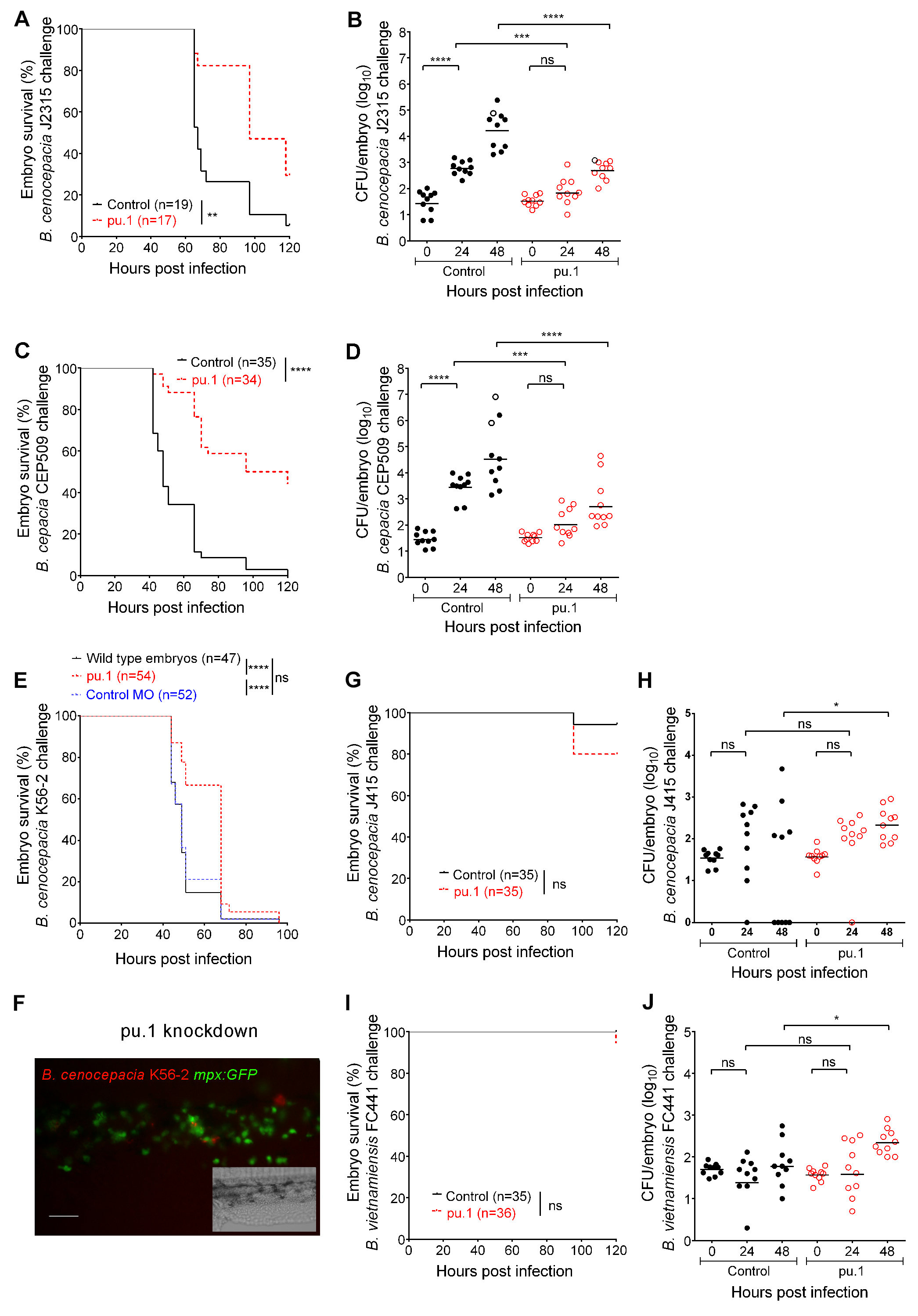
Fig. S1
Related to Figs 1 and 2. Role of macrophages during infection with Bcc strains causing either persistent or acute infection.
(A-D G-J) Embryo survival (A, C, G, and I) and bacterial burden (total of 2 experiments) over time (B, D, H, J) of control (black) and pu.1 knockdown embryos (red) injected iv with B. cenocepacia J2315 (A,B), B. cepacia CEP509 (C,D), B. cenocepacia J415 (G,H), and B. vietnamiensis FC441 (I,J), respectively. (E) Representative experiment (of at least three) showing embryo survival of control embryos (n = 47), pu.1 knockdown (n = 54) and nonspecific control MO (n = 52) embryos injected with B. cenocepacia K56-2 (average 53 CFU). (F) Representative fluorescence image at 24 hpi showing neutrophils (green) in an mpx:GFP pu.1 knockdown embryo injected with B. cenocepacia J2315 (red) (~50 CFU). Inset shows corresponding bright field image. Scale bar, 100 μm. (B, D, H, J) Geometric mean with each data point representing an individual embryo. Dead embryos are indicated as black open circles. (A-E and G-J) * p ≤ 0.05; ** p ≤ 0.01; *** p ≤ 0.001; **** p ≤ 0.0001; ns: non-significant. See materials and methods for statistical tests used.