
|
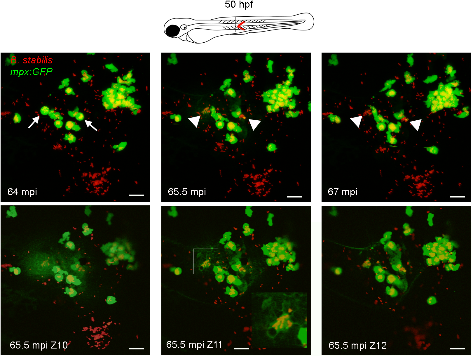
Fig. 5
Neutrophils efficiently phagocytose surface-associated Bcc bacteria.
Confocal stacks after subcutaneous infection with B. stabilis in mpx:GFP embryos (STD intensity projection, 2 μm x 19 steps; T = 35–37 in S2 Movie). Arrows, rounded neutrophils (green) with vacuoles full of bacteria (red) at 64 minutes post injection (mpi) eject their cell contents in the surroundings (arrow heads 65.5 mpi), leaving bacterial clusters and cell debris (arrow heads, 66 min). Lower panels, MAX intensity projection of three consecutive slices (2 μm) at 65.5 mpi showing ejected cellular contents (diffuse GFP signal). Scale bars, 50 μm.