
Fig. 3
- ID
- ZDB-IMAGE-170807-19
- Genes
- Publication
- Passoni et al., 2017 - Imaging of viral neuroinvasion in the zebrafish reveals that Sindbis and chikungunya viruses favour different entry routes
- All Figures
- Figures for Passoni et al., 2017
|
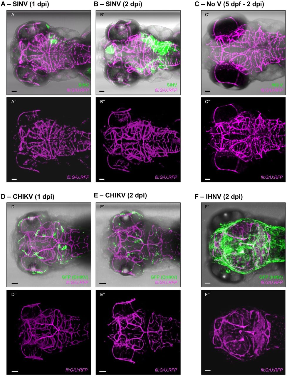
Fig. 3
CHIKV, but not SINV, infects the brain microvascular endothelium. Dorsal views of fli:G/U:RFP larvae, with infected cells in green and endothelial cells shown in magenta; confocal imaging, maximal projections. For all panels, the top image (with ′) shows superposition of transmitted light with green and magenta fluorescence, and the bottom image (with ″) shows only the magenta fluorescence to provide better visualization of the vasculature. Scale bars: 50 μm. (A,B) Live imaging of the same SINV-infected larva at 1 dpi (A) and 2 dpi (B). See also Fig. S1A, Movies 1 and 2, for a view through the z-stack. (C) Live imaging of control uninfected (No V) larva. (D,E) Fixed CHIKV-infected larvae at 1 dpi (D) and 2 dpi (E). See also Fig. S1B and Movie 4, for a view through the z-stack. (F) Fixed IHNV-infected larva at 2 dpi. See also Movie 5, for a view through the z-stack. In this and all following dorsal view figures, anterior is to the left.