
|
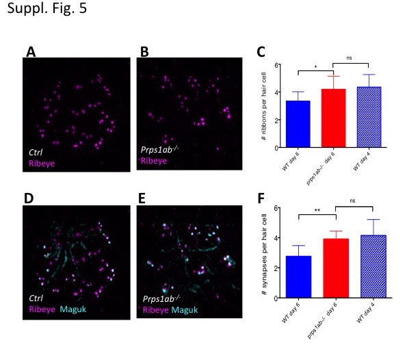
Fig. S5
Analysis of presynaptic ribbons and synapse of neuromast hair cells in the double mutant.
(A-C) Increased presynaptic ribbons in the double mutants. (A, B) Morphology of Ribeye stained presynaptic ribbons of neuromast hair cells in the control (A) and double mutant (B) embryos at 6 dpf. (C) Quantification of the number of presynaptic ribbons in the control and mutant embryos at 6 dpf, as well as in wild-type embryos at 4 dpf. (D-F) Increased synapses in the double mutants. (D,E) Morphology of Maguk stained synapses and Ribeye stained presynaptic ribbons of neuromast hair cells in the control (D) and double mutant (E) at 6 dpf. (F) Quantification of the number of synapses in the control and mutant embryos at 6 dpf, as well as in wild-type embryos at 4 dpf. The numbers of neuromasts analyzed for each staining were 12, 13, and 10 for control, double mutant, and wild type embryos at 4 dpf, respectively.