
|
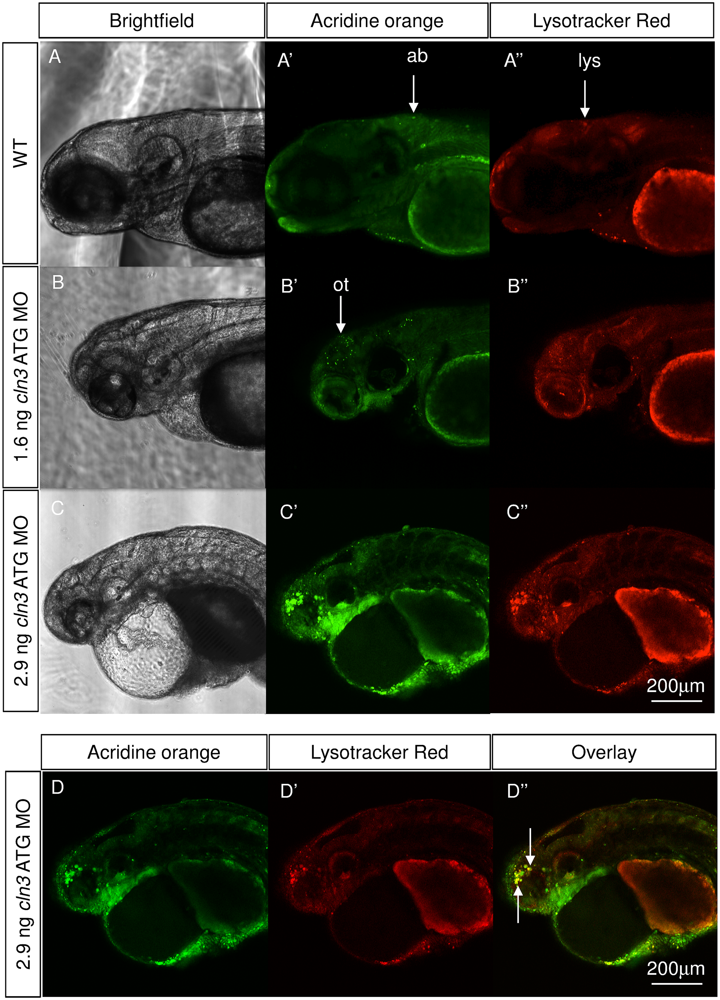
Fig. 8
Apoptotic cells and lysosomal storage were found in the cln3 ATG MO morphant brain.
Acridine orange and Lysotracker staining was carried out on live WT and cln3 ATG MO morphant zebrafish aged 4 dpf. (A-A'') WT fish showed low levels of programmed cell death in the brain and very little bright lysosomal staining. (B-B'') Morphants injected with 1.6 ng of cln3 ATG MO had increased levels of programmed cell death (acridine orange, bright green) in the brain, this localised particularly to the optic tectum, but only a slight increase in lysosomal staining (red). (C-C'') Morphants injected with 2.9 ng of cln3 ATG MO showed very high levels of programmed cell death in the forebrain, midbrain, retina and yolk-body boundary; lysosomal hypertrophy was also observed. (D-D'') Morphants injected with 2.9 ng of cln3 ATG MO exhibit abundant apoptotic bodies in the forebrain that often co-localises with lysosomes (arrows). Bright green, acridine orange in apoptotic bodies; Red, Lysotracker red in lysosomes. Abbreviations: ab, apoptotic body; lys, lysosome; ot, optic tectum. A-A", B-B", C-C'' are Z projections. D-D'' are the same Z slice. Lateral views. Anterior to the left. Dorsal up. The scale bars represent 200 μm and apply to all panels.