
|
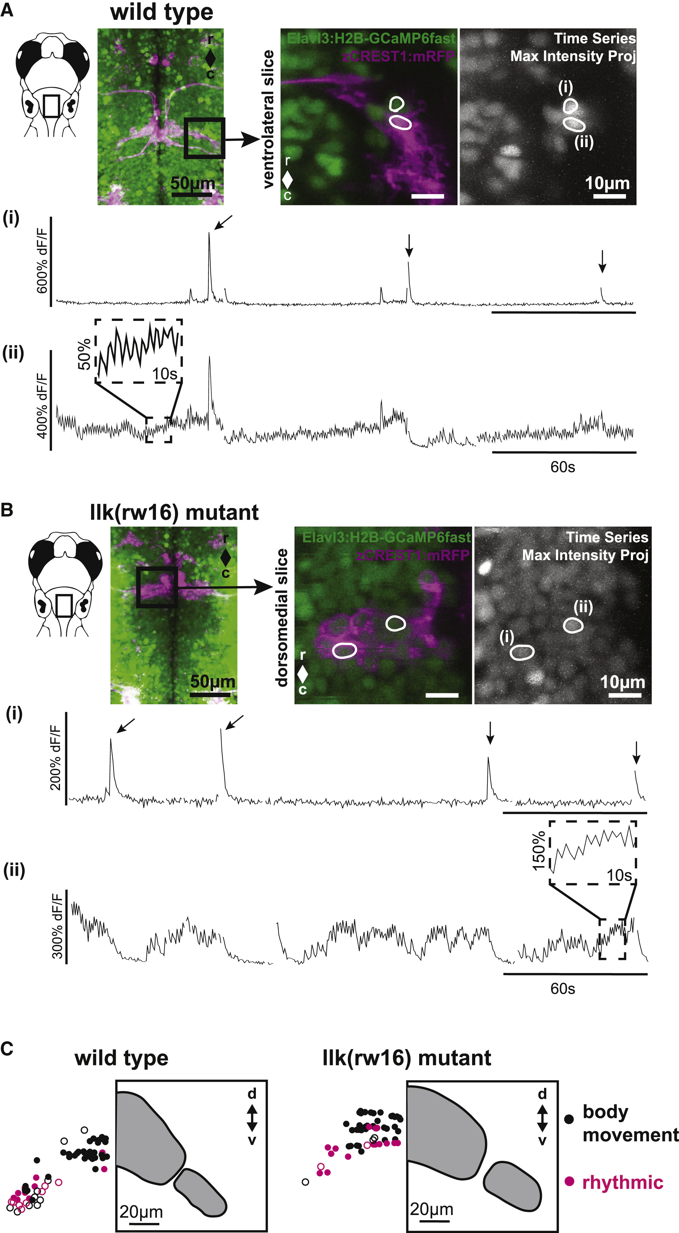
Fig. 6
Calcium Imaging in Intact Larvae Confirms Patterns of FBMN Activity and Reveals a Robust Functional Spatial Topography
(A) In wild-type larvae, some FBMNs exhibit only large, infrequent calcium transients during attempted body movements, whereas other FBMNs exhibit additional rhythmic activity. These two response types could be observed in neighboring FBMNs, as shown here. Outlined regions of interest (i and ii) correspond to the calcium traces shown in (i) and (ii). Sampling interval = 247 ms.
(B) FBMNs in llk(rw16) migration mutants also exhibit these two activity patterns. Sampling interval = 493 ms. Arrows in (A) and (B) indicate large body movements.
(C) In both wild-type (left) and llk(rw16) mutant (right) larvae, there was significant overlap of neurons exhibiting infrequent only (black symbols) and rhythmic (magenta symbols) activity, though rhythmic neurons were concentrated ventrolaterally (Table S2). Open symbols, neurons backfilled from the LO (operculum). Grey panels show the cross-sectional outline of the facial motor nucleus for each phenotype, based on backfill data shown in Figure 2. Number of fish used to assemble summary data: n = 7 wild-type (64 classified neurons in total); n = 6 llk(rw16) mutant (60 classified neurons in total). All data shown here were obtained from unparalyzed fish at 5 dpf.