
|
Fig. 4
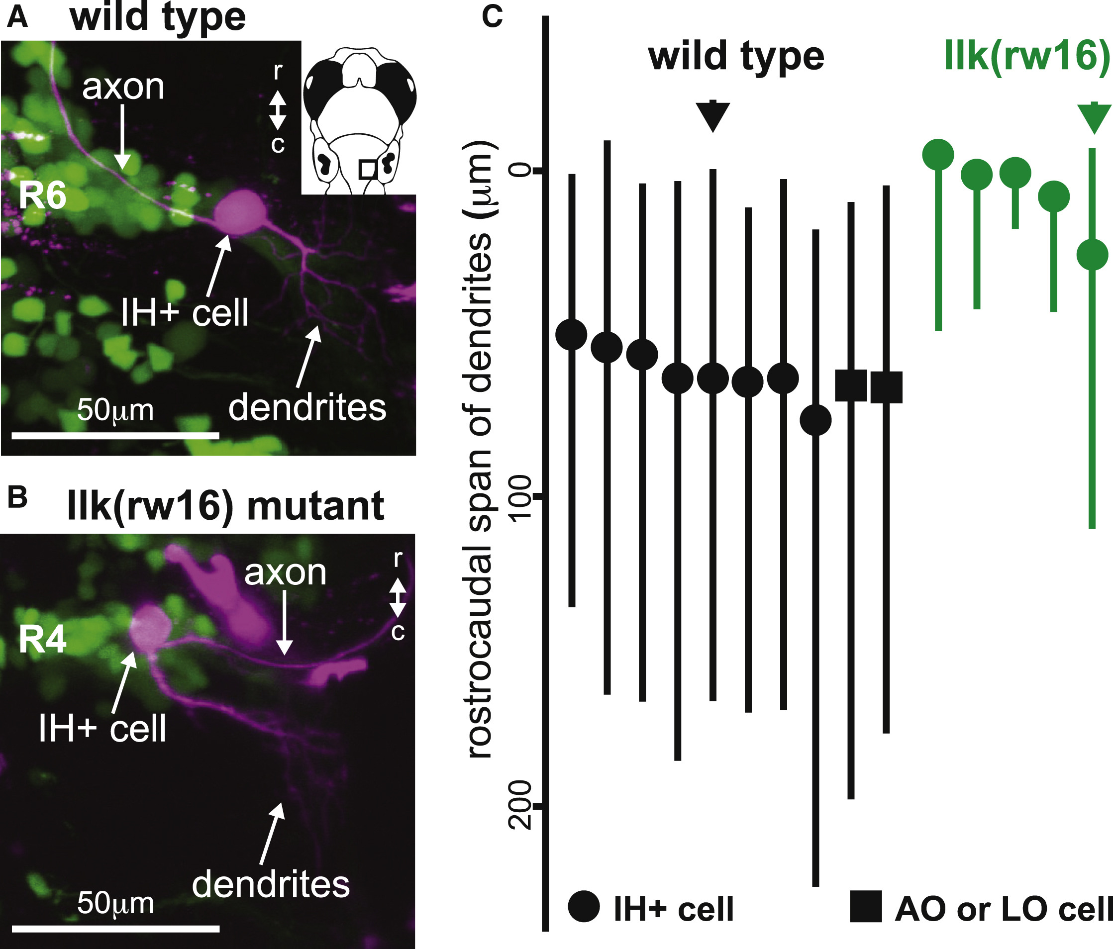
The Location of Dendrites from Facial Motor Neurons in Wild-Type and Migration Mutants
(A and B) In both wild-type (A) and llk(rw16) mutant (B) larvae, single-cell dye fills show that FBMNs extend their dendrites primarily into the neuropil caudal and lateral to the cell body.
(C) However, a comparison of the rostrocaudal span of FBMN dendrites reveals that wild-type FBMNs innervate more caudal regions of the neuropil than migration mutant FBMNs.
Circles, IH+-projecting FBMNs; squares, operculum-projecting FBMNs. Black and green arrows correspond to the neurons used in (A) and (B), respectively. All cells were filled at 4 dpf and imaged at 5 dpf.