
|
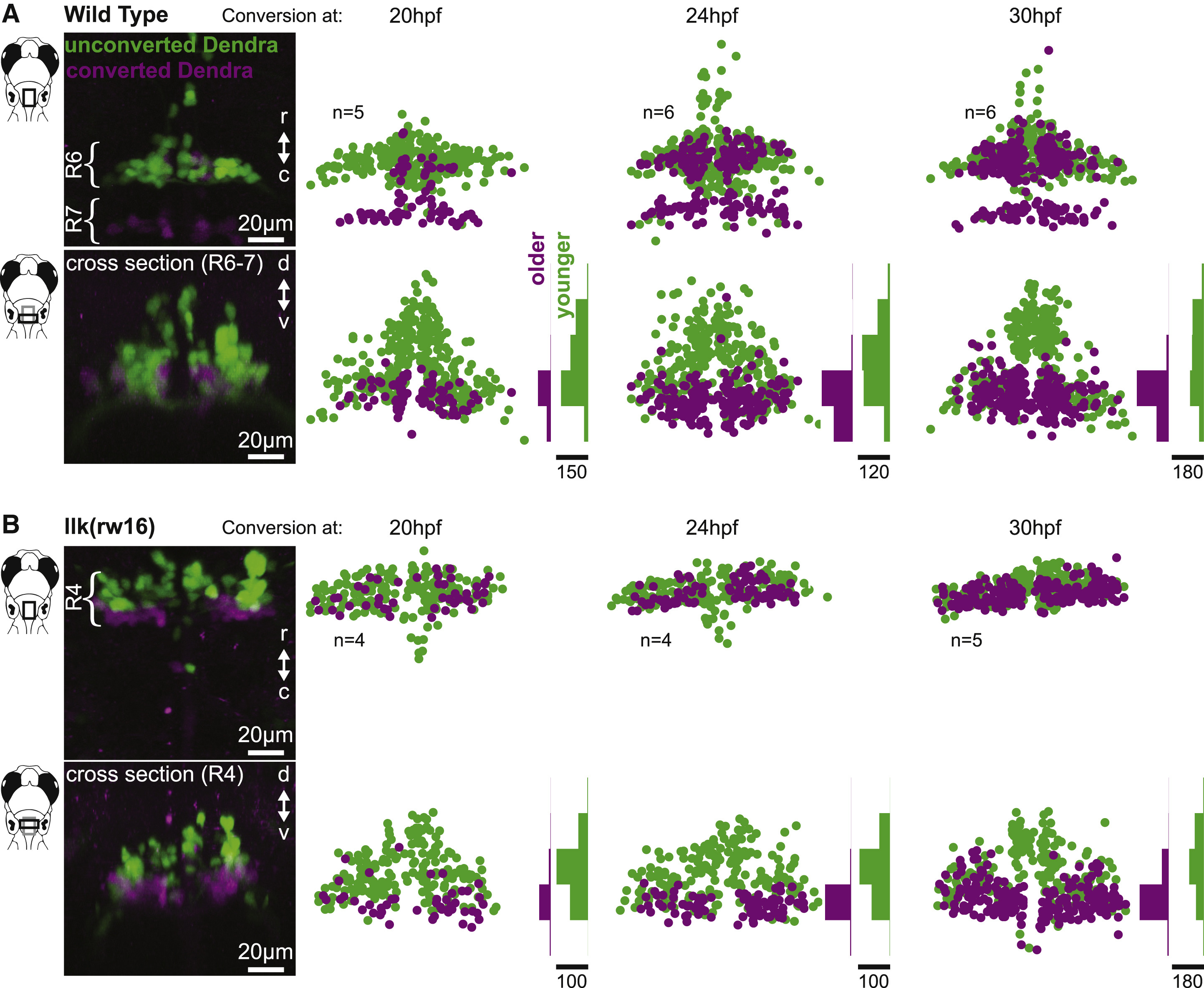
Fig. 3
Facial Motor Neurons Are Topographically Organized According to Age in Both Wild-Type and Caudal Migration Mutants
Zebrafish embryos expressed the photo-convertible protein Dendra2 in cranial motor neurons and were exposed to UV light at one of three time points during facial motor neuron differentiation (20, 24, or 30 hpf) and then imaged at 3 dpf. Neurons older than the conversion time point contain converted (magenta/purple) Dendra, whereas younger neurons contain only unconverted (green) Dendra. In both wild-type (A) and llk(rw16) mutant (B) larvae, older facial motor neurons are located in the ventral-most part of the facial motor nucleus. Examples in left panels of (A) and (B) have been color adjusted to increase visibility of the converted (magenta) Dendra signal. Histograms illustrate the distribution of cells along the dorsoventral axis binned at 20-μm intervals, where the scale bars refer to the number of cells. See Figure S2 for data from pk1b(fh122) mutants.