
|
Fig. 1
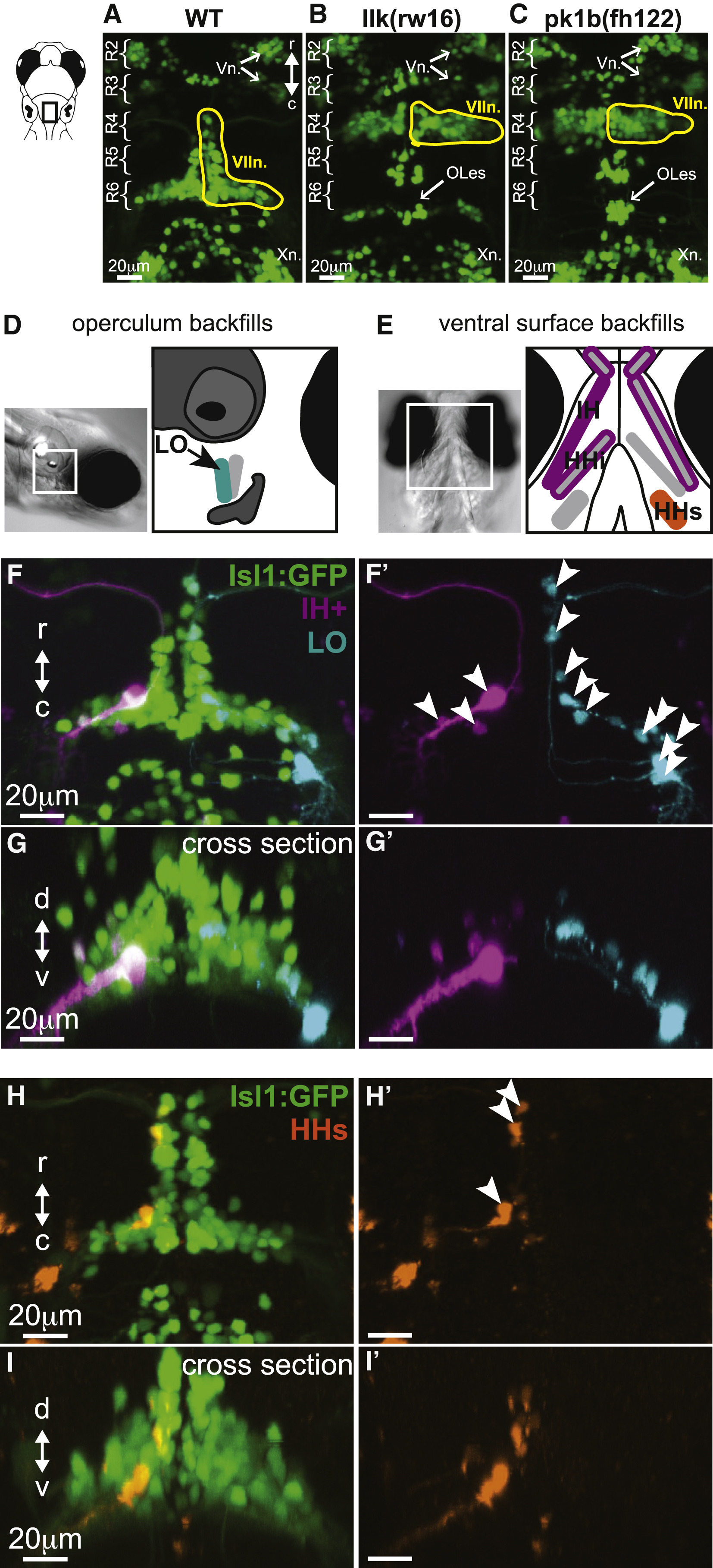
Facial Motor Neurons Migrate Caudally in Wild-Type, but Not Migration Mutant, Larvae, and Backfills from Cranial Muscles Reveal the Location of Facial Motor Pools
Top panels show the hindbrain of Tg(Islet1:GFP) larvae at 5 dpf.
(A) In wild-type larvae, facial motor neurons are born in rhombomere 4 (R4) and migrate caudally to settle in rhombomeres 5–7 by 48 hpf.
(B and C) In both llk(rw16) mutant larvae (B) and pk1b(fh122) mutant larvae (C), facial motor neurons fail to migrate caudally and remain in R4. Octavolateralis efferents (OLes) born in R4 and R6 also fail to migrate caudally.
(D and E) Schematic representation of the operculum (D) and ventral cranial muscles (E) targeted for backfills of the facial motor nerve. Filled muscles indicate the electroporation target, and outlined muscles correspond to other muscles labeled with dye after target electroporation.
(F and G) Electroporation of fluorescent dyes into the IH (magenta) and LO (cyan) muscles on opposite sides of a single zebrafish larva (4 dpf) backfills a subset of each muscle's facial motor pool, imaged at 5 dpf, and shown here from dorsal (F and F') and cross-sectional (G and G') views.
(H and I) Targeting the electroporation to the HHs (orange) backfills a subset of that muscle's motor pool, shown here from dorsal (H and H') and cross-sectional (I and I') views.
Arrowheads indicate backfilled cell bodies. HHs, hyohyoideus superior; IH+, interhyoideus + hyohyoideus inferior; LO, levator operculi; Vn., trigeminal motor nuclei; VIIn., facial motor nucleus; Xn., vagal motor nucleus. See also Figure S1.