
Fig. 7
- ID
- ZDB-IMAGE-170620-9
- Publication
- Miller et al., 2017 - A genetic basis for molecular asymmetry at vertebrate electrical synapses
- All Figures
- Figures for Miller et al., 2017
|
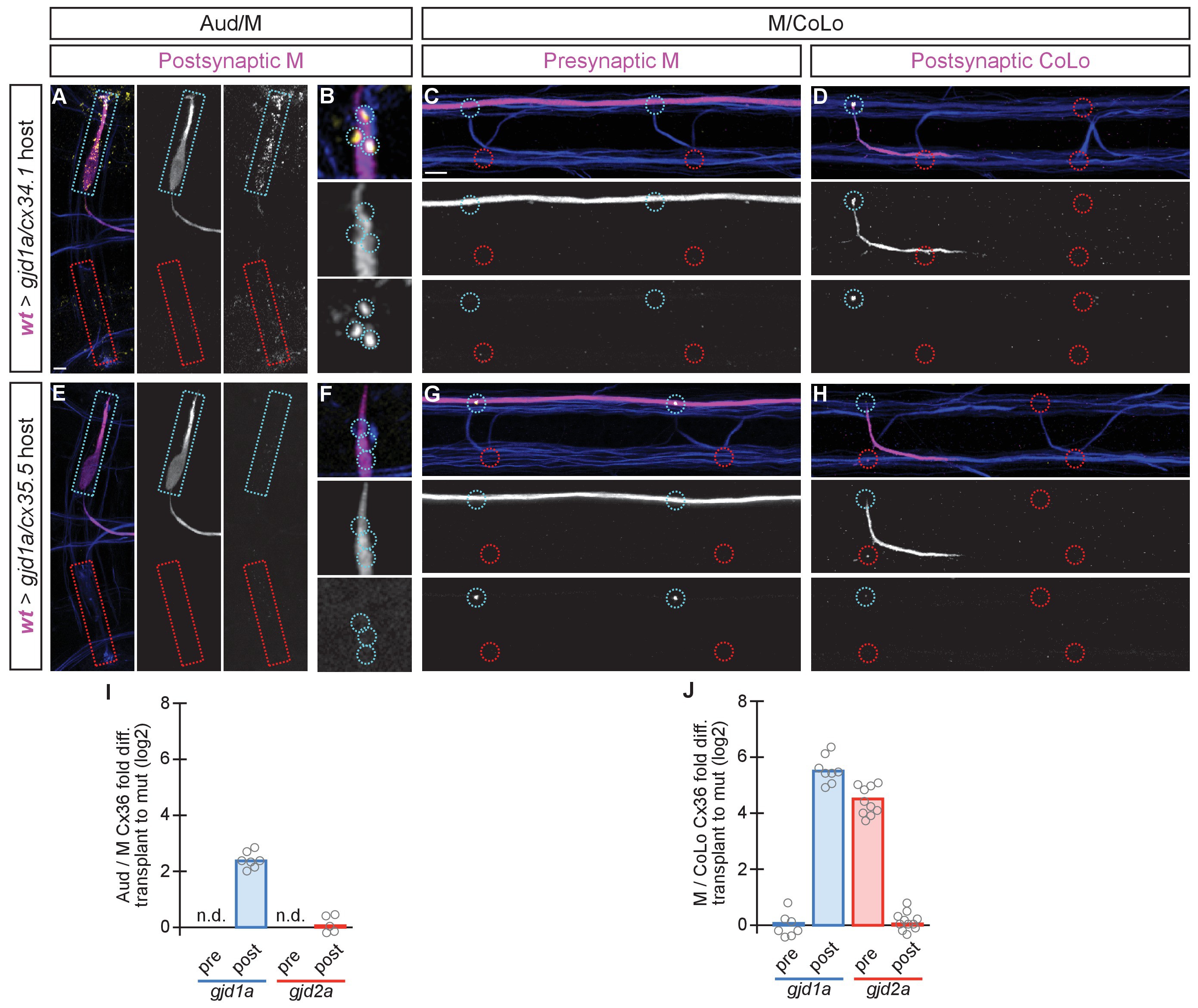
Fig. 7
The exclusive asymmetric functions of gjd1a/cx34.1 and gjd2a/cx35.5 are sufficient for electrical synapse formation.
Dorsal views of chimeric larvae containing GFP-marked cells transplanted from a wildtype (wt) donor embryo into a mutant host of noted genotype; throughout the figure the neurons derived from the wt donor embryo are displayed in magenta, while those from the mutant host are in blue. Synapses (stained with anti-human-Cx36, yellow) associated with a transplanted neuron (cyan circles and boxes) can be directly compared to mutant host synapses (red circles and boxes). Hindbrain, lateral dendrite, and spinal cord images are maximum intensity projections of ~30, ~5, and ~10 uM, respectively. Anterior is to the left. Scale bar = 10 uM. (A–D) When the postsynaptic neuron of the Aud/M or M/CoLo synapse is wildtype in a gjd1a/cx34.1 mutant the electrical synapses are rescued (A,B,D). By contrast, when the presynaptic neuron is wildtype in a mutant there is no effect on Cx36 staining at the synapse (C). (E–H) Conversely, electrical synapses are rescued in gjd2a/cx35.5 mutants only when the presynaptic, and not the postsynaptic, neuron is wildtype. (I,J) Quantitation of the fold increase in the ratio of Cx36 at wt-donor-associated synapses to mutant host synapses in chimaeric embryos. Each circle represents the average ratio of 1–8 donor-associated to 8–16 host-associated synapses within an animal. Associated quantitation of Cx36 at chimeric synapses can be found in Figure 7—source data 1 for Figure 7 and Figure 7—source data 2 for Figure 7.