
|
Fig. 5 S1
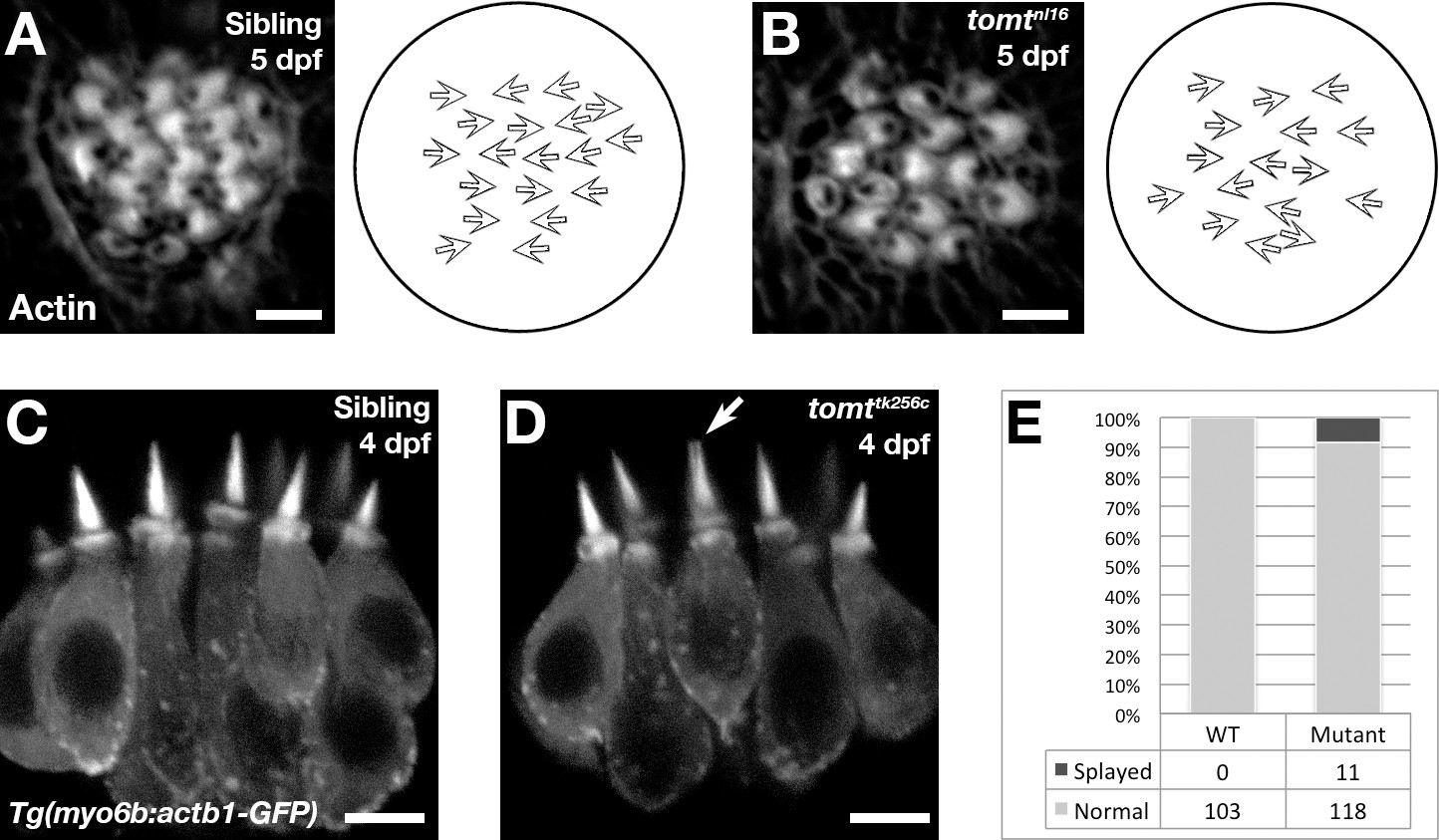
Hair bundle polarity and morphology in Tomt-deficient hair cells.
(A, B) Hair cell planar polarity in neuromasts of mercury mutants. Images of hair bundles from 5 dpf wild-type sibling (B) and tomtnl16 mutant (C) larvae stained with phalloidin-488 for F-Actin. Accompanying each image is a diagram of the planar polarity of each hair bundle. (C–E) Inner ear hair cell morphology in mercury mutants. (C, D) Representative images of hair cells in lateral cristae expressing βactin-GFP from Tg(myo6b:actb1-GFP) wild-type siblings (D) and tomttk256c mutants (E) at 4 dpf. White arrow indicates a splayed hair bundle sometimes observed in mercury mutants. (E) Quantification of the splayed bundle phenotype in wild-type siblings (n = 6 larvae, 103 lateral crista hair bundles), and tomttk256c mutants (n = 8 larvae, 129 lateral crista hair bundles). Scale bars = 3 µm in B and C, 5 µm in D and E.