
|
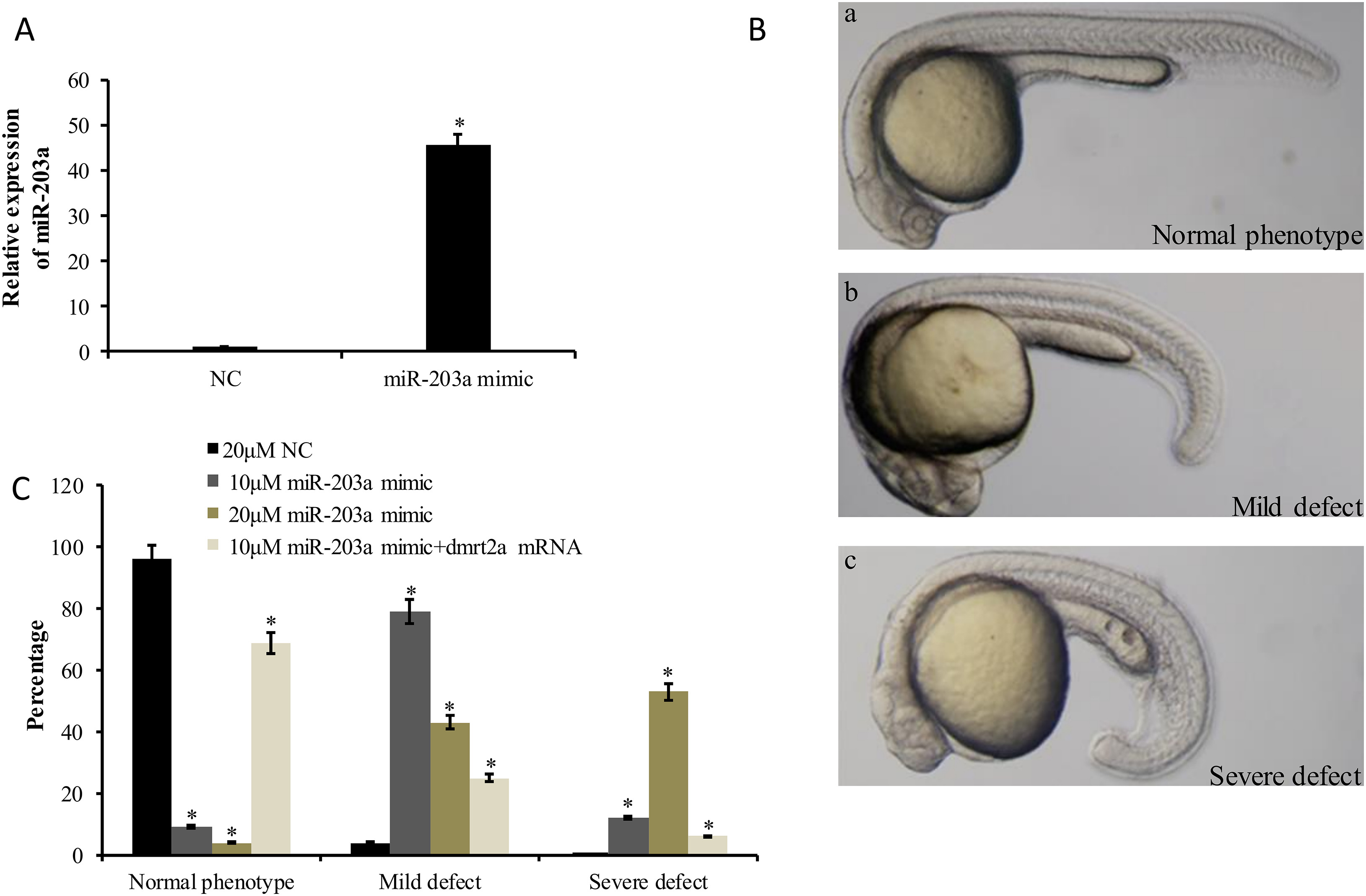
Fig. 4
Morphological observation and statistical analysis for miR-203a mimic and its negative control mimic (NC) injected group at 24 hpf. (A) Levels of miR-203a in 24 hpf embryos injected with NC and miR-203a mimic. 30 embryos in each experiment. (B) The morphology of normal phenotype embryos (a), mild defect embryos (b) and severe defect embryos (c) after injection. (C) The percentage of normal phenotype, mild defect and severe defect in 20 μM NC, 10 μM and 20 μM miR-203a mimic, 10 μM miR-203a mimic and dmrt2a mRNA injected embryos. 100 embryos in each experiment. Error bars indicate mean ± SD, n = 3. Student's t-test was used for statistical analysis (*P < 0.05).
Reprinted from Gene, 625, Lu, C., Wu, J., Xiong, S., Zhang, X., Zhang, J., Mei, J., MicroRNA-203a regulates fast muscle differentiation by targeting dmrt2a in zebrafish embryos, 49-54, Copyright (2017) with permission from Elsevier. Full text @ Gene