
|
Fig. S8
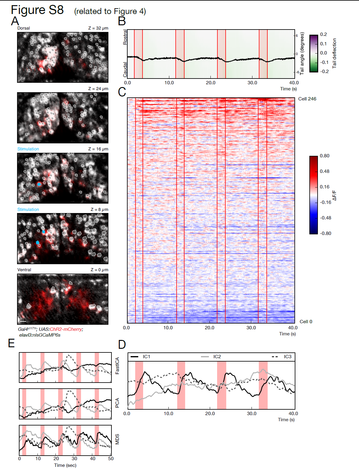
Investigation protocol demonstrated in an additional larva (related to Figure 4).
(A) Volumetric recording of the activity of hundreds of neurons during stimulation-induced behavior. 5 planes are imaged in the midbrain of a fish expressing ChR2-mcherry (red) and nlsGCaMP6s (grey). ROIs corresponding to the cell bodies selected for the analysis are highlighted in white, and neurons selected for the photostimulation are shown in cyan.
(B) The kinematics of the tail show a reliable tail deflection upon stimulation (red lines indicate stimulation onset and offset).
(C) Raster plot showing the activity for the recorded population of 246 neurons, temporally aligned with the behavior recording shown in B. Similar to the example in Figure 4, a small subset of neurons shows a reliable activity pattern temporally locked to the stimulation and tail deflection.
(D) Representation of common patterns in population activity by independent component analysis. The first component captures the stimulation induced network activity that corresponds to the tail bending behavior.
(E) Comparison of dimensionality reduction methods on the same data shown in Figure 4. The different methods tested, Independent Component Analysis (fastICA), Principal Component Analysis (PCA), and Multi-Dimensional Scaling (MDS), all capture a rather similar representation of the network. One component captures the consistent activity induced by photostimulation, and a different component picks the activity associated with the large swim. The ICA and PCA representations are almost identical, while for MDS, which is a non-linear a manifold learning technique, the activity induced by stimulation is partially distributed into two components. Scale bar is 10 μm.