
|
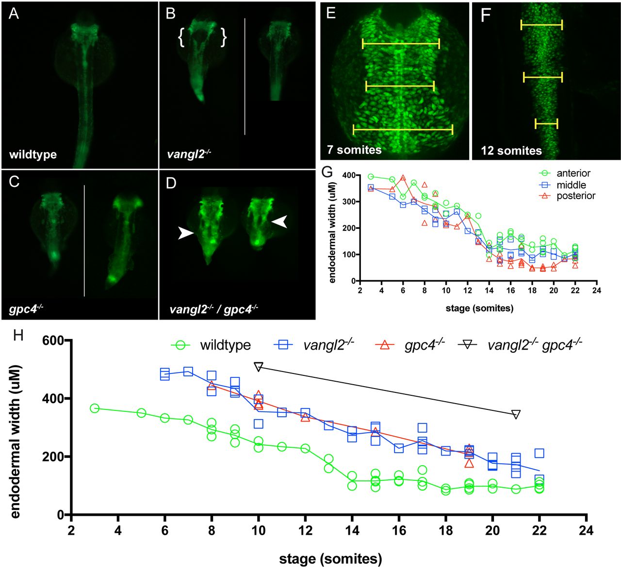
Fig. 2
Loss of PCP signalling results in disrupted endoderm morphology during development. (A-D) Fluorescent dissecting microscope images of endoderm morphology at 24 hpf. All views are dorsal. (A) Wild-type and PCP homozygous mutant embryos (B) vangl2, (C) gpc4, and (D) vangl2l/gpc4 double mutants in the Tg(sox17:EGFP) background. Endoderm morphology in PCP mutants (B, C and D) is disorganised and wider than in wild-type (A). Disruption of PCP signalling in the single mutants occasionally results in a splitting of the endoderm posterior to the pharyngeal endoderm as indicated by the brackets in B. vangl2/gpc4 double mutant embryos always have a splitting of the endoderm posterior to the pharyngeal endoderm as indicated by the arrow heads in D. (E-F) Confocal projections of 7 somite (E) and 12 somite (F) wild-type Tg(sox17:EGFP) embryos. Dorsal views. Yellow bars indicate positions of the three width measurements. (G) The rate of change of the width of the endoderm, measured at anterior, middle, and posterior regions of the trunk endoderm, migrate with the same overall rate towards the dorsal midline during somitogenesis stages. (H) The endoderm is wider in homozygote vangl2 and gpc4 mutants and vangl2/gpc4 double mutant lines, although dorsal migration does still take place (n = 1-5 embryos per stage, per genotype). White lines in panel C and E divide grouped individual embryos.