
|
Fig. 5 S1
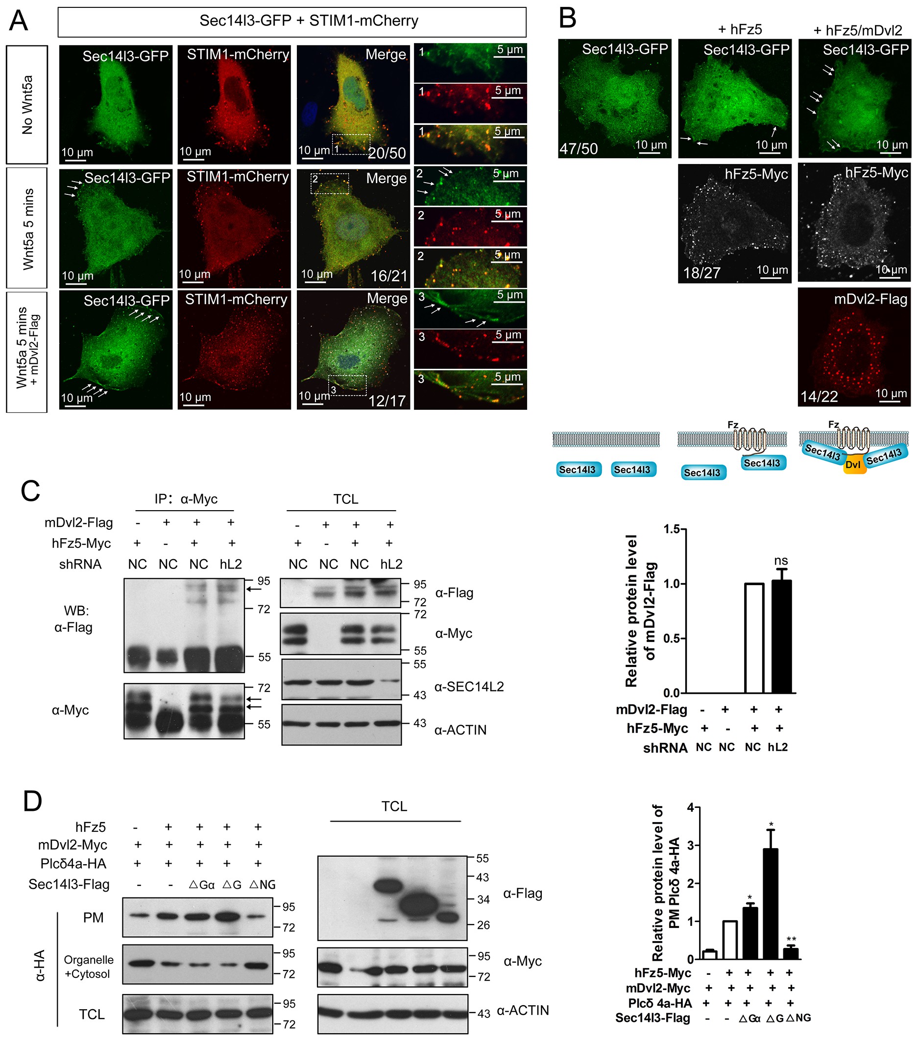
The subcellular localization of Sec14l3 and its function in the Plcδ4a PM recruitment.
(A) Co-localization of highly expressed Sec14l3 with STIM1 proteins in PC3 cells with or without Wnt5a stimulation. The three panels show the localization of Sec14l3 and STIM1 without Wnt5a stimulation, with 400 ng/μl Wnt5a stimulation for 5mins, and Wnt5a stimulation with mDvl2 co-transfection respectively. Arrowheads indicate PM-localized Sec14l3-GFP and STIM1-mCherry. Scale bars, 10 μm. (B) Sec14l3 was recruited to the PM in the presence of hFz5 and mDvl2. Arrowheads indicate PM-localized Sec14l3-GFP. Scale bars, 10 μm. (C) The interaction between hFz5 and mDvl2 is not affected after SEC14L2 knockdown in HEK293T cells. Quantification of mDvl2 protein levels is shown on the right (see also Figure 5—source data 1, ns, non-significant, p>0.05). (D) Plcδ4a recruitment to the PM induced by hFz5 overexpression is inhibited byΔNG form of Sec14l3 in the PC3 cells. Quantification of PM Plcδ4a protein levels is shown on the right (see also Figure 5—source data 1, **p<0.01; *p<0.05).