
|
Fig. 9 S1
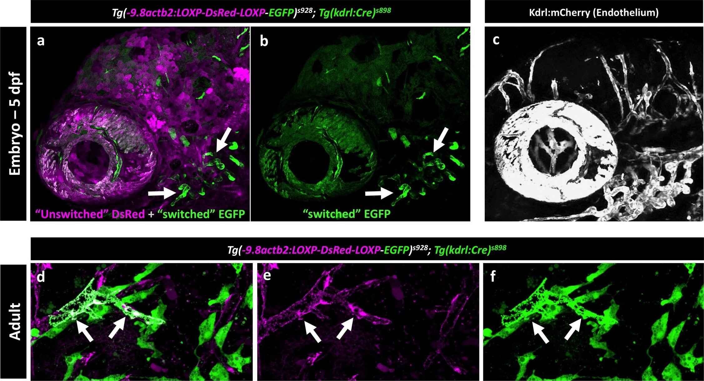
Mosaic expression of EGFP in vessel endothelial cells in the Kdrl:Cre ‘switch’ double transgenic line
(a,b) Lateral view confocal micrograph of the head of a 5 dpf Tg(−9.8actb2:LOXP-DsRED-LOXP-EGFP); Tg(kdrl:Cre) double transgenic ‘switch’ animal, showing DsRed-positive ‘unswitched’ cells (magenta, panel a) and mosaic EGFP expression in only a subset of ‘switched’ endothelial cells (green, panels a and b; arrows note switched EGFP positive endothelium). (c) Lateral view confocal micrograph of the head of a control 5 dpf Tg(kdrl:mCherry) animal, showing uniform mCherry transgene expression (grey) in all endothelial cells. (d-f) Higher magnification confocal micrographs of the same adult brain shown in Figure 9f, with arrows noting a partially switched vessel segment that is both DsRed positive (magenta, panels d and e) and EGFP positive (green, panels d and f).
ne.