
Image
|
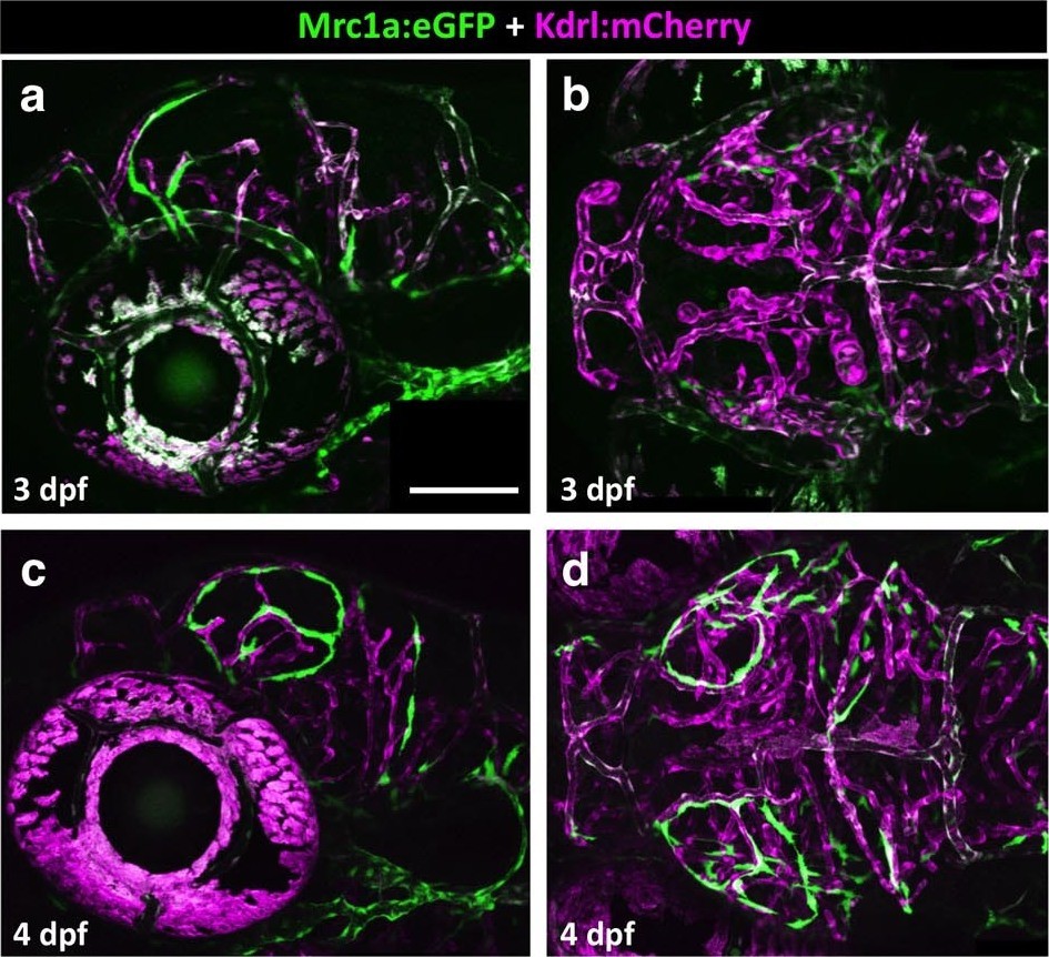
Figure Caption
Fig. 7 S1
Emerging FGPs on the 3 and 4 dpf zebrafish brain.
(a-d) Confocal images of Mrc1a:eGFP-positive FGPs (green) and Kdrl:mCherry-positive blood vessels (magenta) on the surface of the brain in 3 dpf (a,b) and 4 dpf (c,d) Tg(mrc1a:eGFP);Tg(kdrl:mCherry) double-transgenic zebrafish. Panels a and c are lateral views, panels b and d are dorsal views of the head. Images are the unmanipulated versions of the images shown in Figure 7a–d, where some residual blood vessel GFP fluorescence was deleted for clarity. Rostral is to the left in all panels. Scale bars = 100 µm. Rostral is to the left. Scale bar = 200 μm.
Acknowledgments
This image is the copyrighted work of the attributed author or publisher, and
ZFIN has permission only to display this image to its users.
Additional permissions should be obtained from the applicable author or publisher of the image.
Full text @ Elife