
|
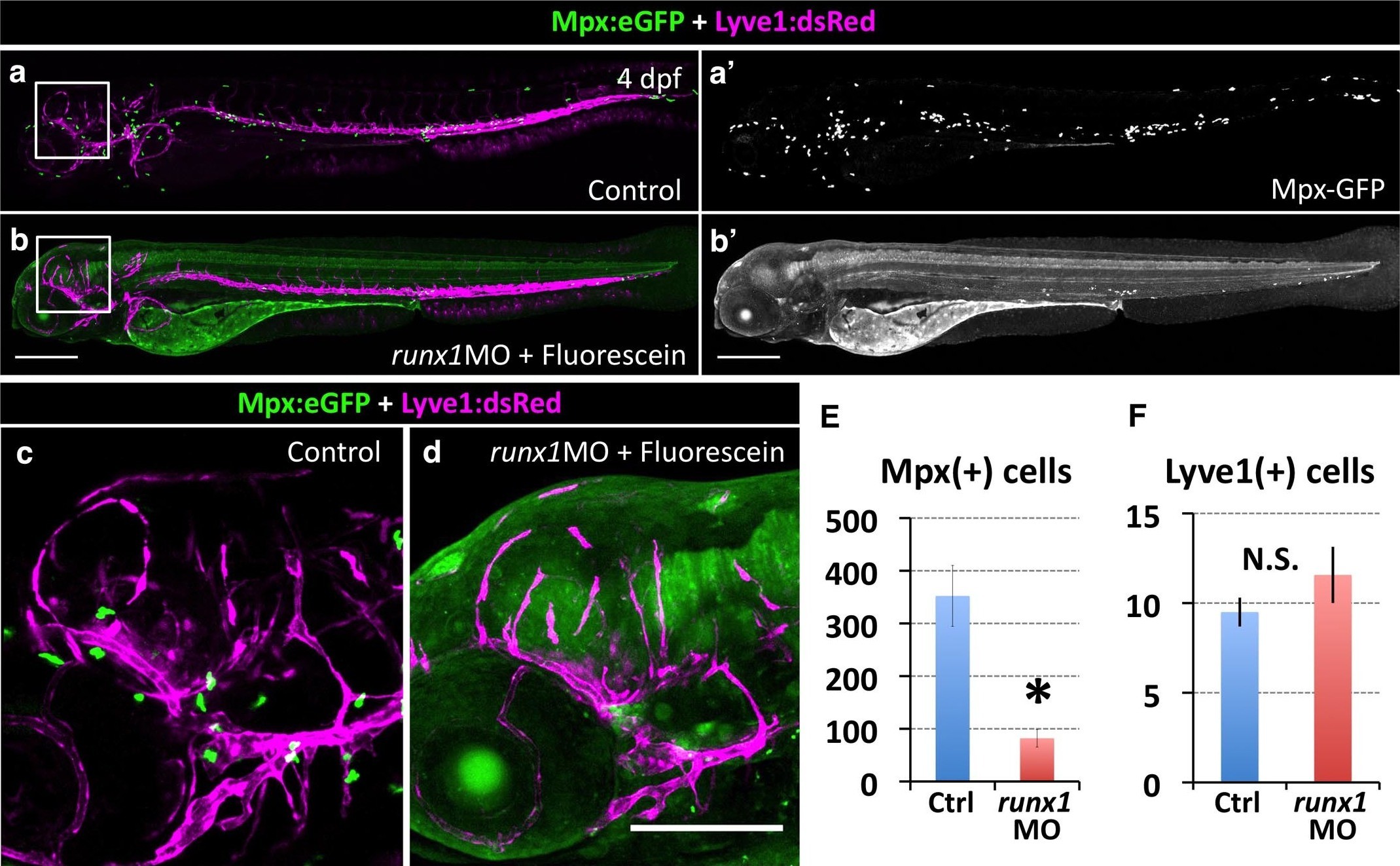
Fig. 5 S3
Inhibiting HSPCs specification does not affect FGP formation.
(a,a’,b,b’) Lateral views of 5 dpf Tg(lyve1:dsRed);Tg(mpx:eGFP) double transgenic animals showing the lymphatic vessels and FGPs (magenta) and macrophages (green punctate) in an uninjected (a) or runx1 Fluorescein-tagged morpholino injected (b) animal. Panels a’ and b’ show only the green fluorescence channel. (c,d) Higher magnification lateral views of the boxed regions in panels a and b, respectively, showing Lyve1:dsRed-positive FGPs localized to the optic tectum at 5 dpf. (e,f) Quantification of Mpx:eGFP-positive cells (e, n = 4 animals imaged and quantitated) and Lyve1:dsRed-positive FGPs (f, n = 8 animals imaged and quantitated) in 5 dpf Tg(lyve1:dsRed);Tg(mpx:eGFP) double transgenic animals after runx1 morpholino injection. Scale bars in all panels: 200 µm.