
Image
|
Figure Caption
Fig. 1 S1
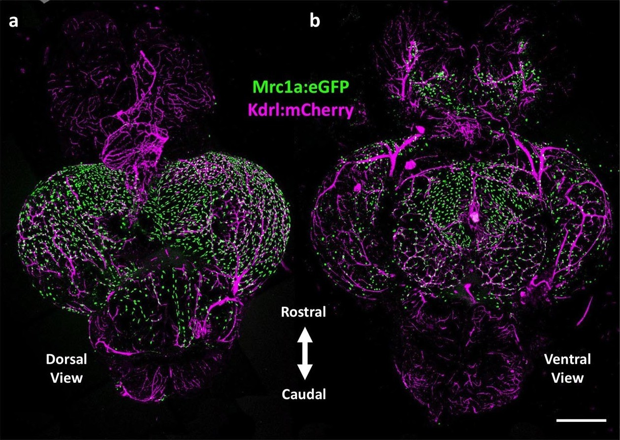
Mrc1a-positive perivascular cells cover the zebrafish brain.
(a,b) Dorsal (a) and ventral (b) confocal images of the dissected brain of a Tg(mrc1a:eGFP);Tg(kdrl:mCherry) double-transgenic adult zebrafish (eGFP and mCherry are shown in green and magenta, respectively). Rostral is to the top. Mrc1a:eGFP cells populate the entire optic tectum on the dorsal and ventral sides but only the ventral side of the forebrain/telencephalon and the dorsal side of the cerebellum. Scale bar: 500 µm.
Acknowledgments
This image is the copyrighted work of the attributed author or publisher, and
ZFIN has permission only to display this image to its users.
Additional permissions should be obtained from the applicable author or publisher of the image.
Full text @ Elife