
Image
|
Figure Caption
Fig. 5
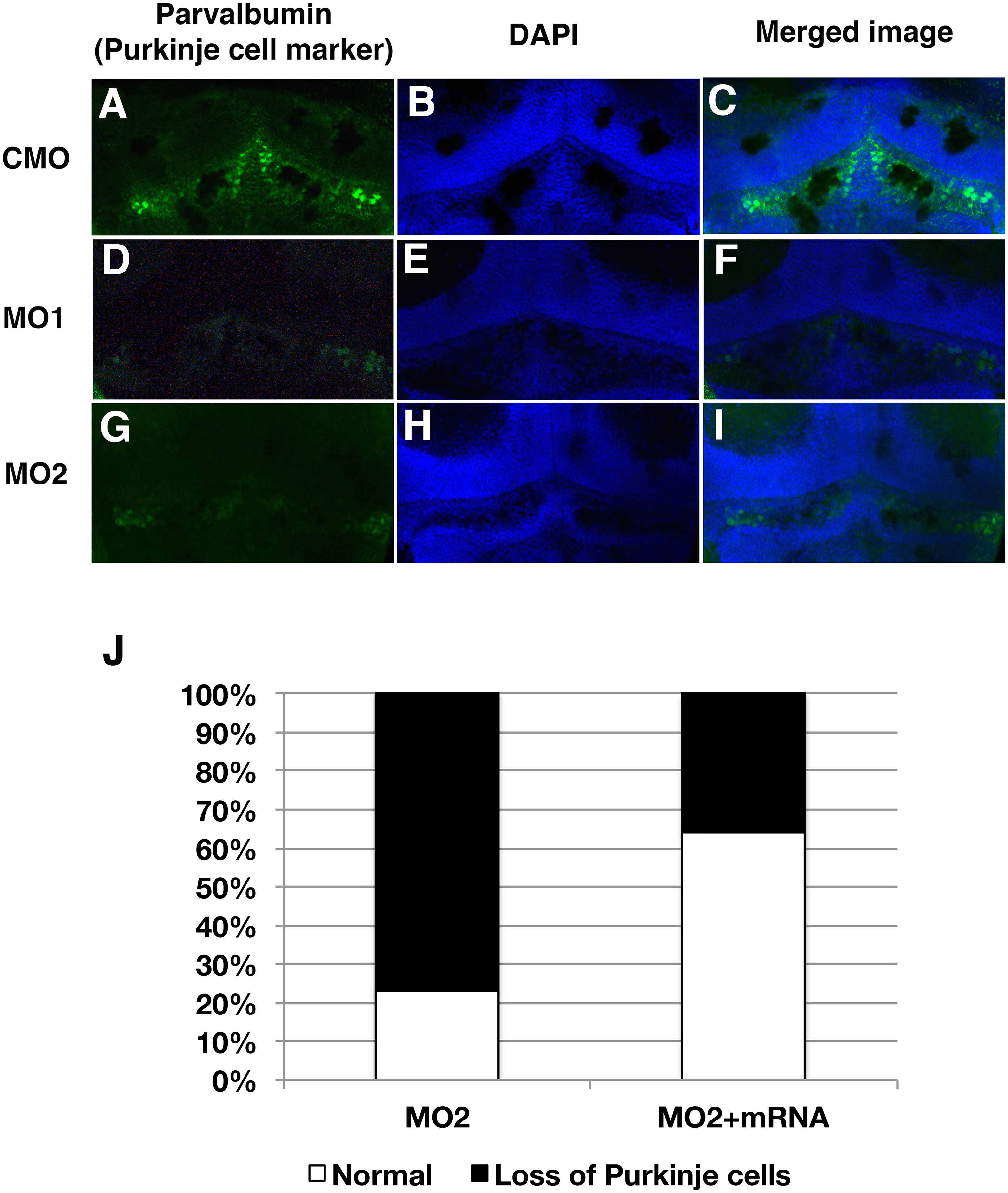
Staining of purkinje cells with anti-purvalbumin in cerebellar area.
The number of purkinje cells detected with anti-parvalbumin shows reduced number of positive cells in MO1 or MO2 injected embryos. (A, B and C): CMO injected fish, (D, E and F): MO1 injected fish, (G, H and I): MO2 injected fish. (A, D and G: Staining Purkinje cell. (B, E and H): DAPI, nuclei. (C, F and I): Merged images. (J): Increased number of Purkinje cells in sil1 morphants with co-injection of fish sil1 mRNA (50 pg) from 23.1% (MO2, n = 13) to 64.3% (MO2+sil1 mRNA, n = 14).
Figure Data
Acknowledgments
This image is the copyrighted work of the attributed author or publisher, and
ZFIN has permission only to display this image to its users.
Additional permissions should be obtained from the applicable author or publisher of the image.
Full text @ PLoS One