
|
Fig. S3
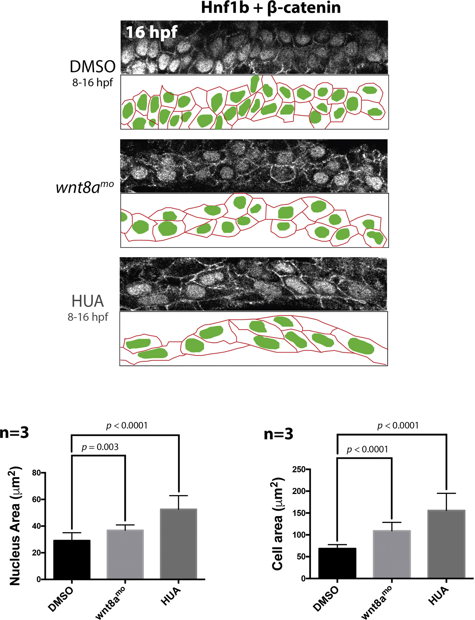
A low dose of wnt8a morpholino and HUA increase the size of pronephric cells and nuclei. Top panels show co-labelling for Hnf1b and β-catenin at 16 hpf after the indicated treatments. Views are dorsal images of the proximal tubule, with lower panels showing schematics of the outlines of pronephric cells and nuclei from the image above. Histograms show nuclear and cell sizes (μm2) of 10 proximal tubule cells in three different animals. Comparing the ratio of the lengths of the AP and DV axes of the cells in each treatment (in μms), we find that when 10 cells in three different embryos were measured, the AP:DV in un-injected controls is 1:1.2. In wnt8a morphants the AP:DV ratio is 1.6:1, and in HUA treated animals it is 2.9:1. These results demonstrate the cells are stretched in wnt8a depleted and HUA treated animals relative to controls.
Reprinted from Developmental Biology, 425(2), Naylor, R.W., Han, H.I., Hukriede, N.A., Davidson, A.J., Wnt8a expands the pool of embryonic kidney progenitors in zebrafish, 130-141, Copyright (2017) with permission from Elsevier. Full text @ Dev. Biol.