
|
Fig. S4
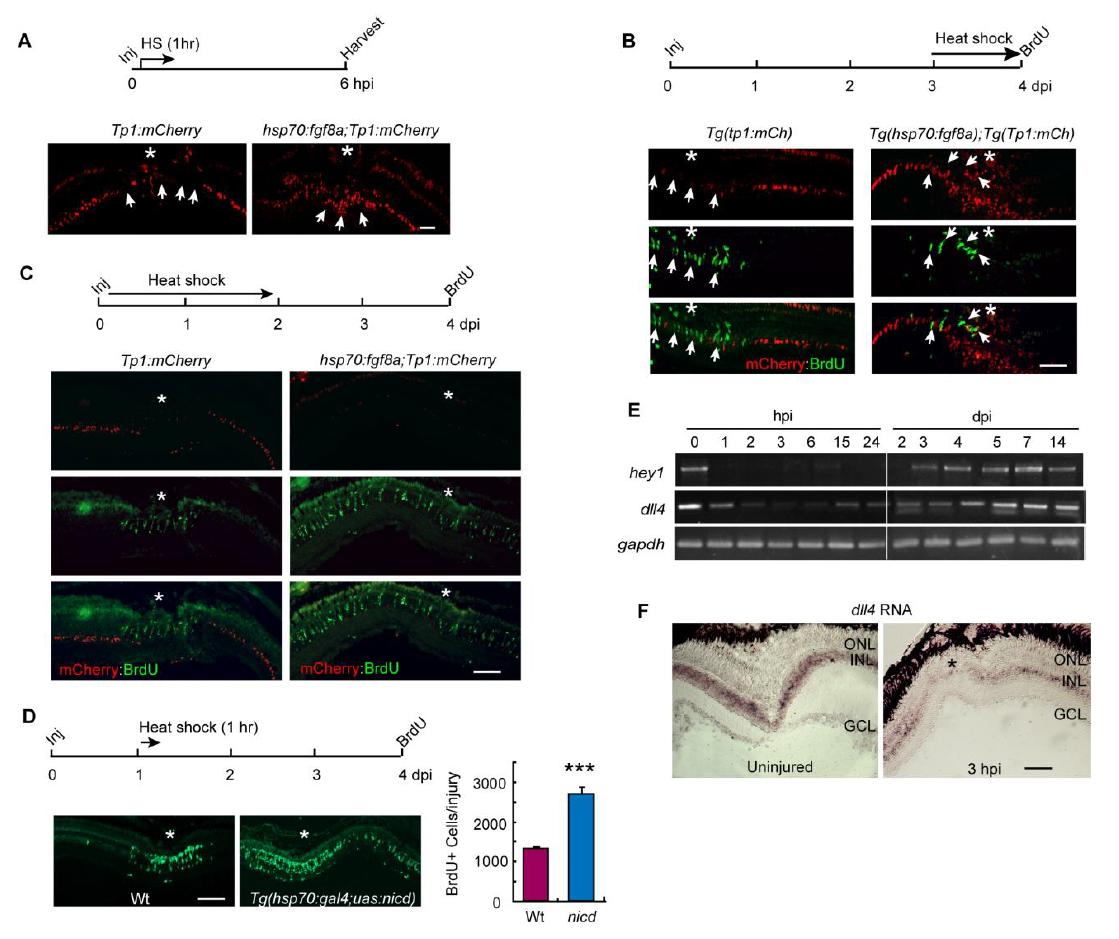
Related to Figure 4. Fgf8a stimulates Notch signaling in the injured retina.
(A) mCherry fluorescence in injured retina from Tp1:mCherry and hsp70:fgf8a;tp1:mCherry fish that received a 1h HS and then sacrificed 5h later when Fgf8a levels are still high. Asterisk and arrows mark the injury site (central retina, 6 mo old fish); scale bar is 100 µm.
(B) mCherry and BrdU immunofluorescence in injured retinas from Tp1:mCherry and hsp70:fgf8a;tp1:mCherry fish that received HS from 3-4 dpi and sacrificed at 4 dpi. Asterisks and arrows mark the injury site (central retina, 6 mo old fish) and arrows point to BrdU+ cells; scale bar is 100 µm.
(C) mCherry fluorescence and BrdU immunofluorescence in injured retina from Tp1:mCherry and hsp70:fgf8a;tp1:mCherry fish that received a 2 day HS and then sacrificed 2 days later. Asterisk and arrows mark the injury site (central retina, 6 mo old fish); scale bar is 100 µm.
(D) BrdU immunofluorescence in injured retinas from Wt and hsp70:gal4:uas:NICD fish that received HS for 1h at 1 dpi and sacrificed at 4 dpi. Asterisk indicates injury site (central retina, 6 mo old fish); scale bar is 100 µm. Graph is quantification of BrdU+ cells/injury; n=3 individual experiments, error bars are s. d. ***P<0.001.
(E) RT-PCR analysis of hey1 and dll4 gene expression in uninjured and injured retina.
(F) In situ hybridization reveals spatial changes in dll4 expression in the injured retina. Asterisk indicates injury site (central retina, 6 mo old fish). ONL, outer nuclear layer; INL, inner nuclear layer; GCL, ganglion cell layer.