
|
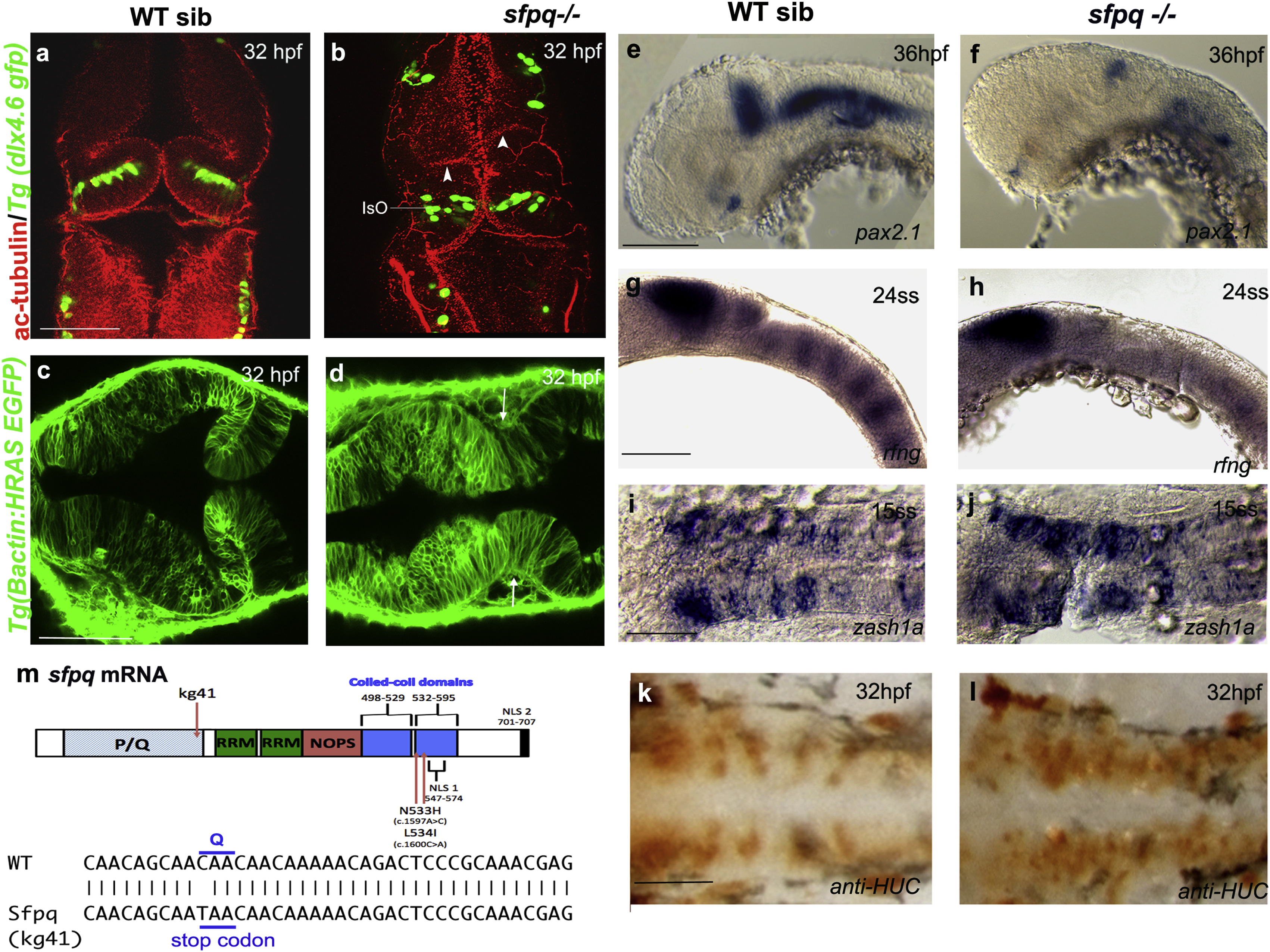
Fig. 1
SFPQ Is Required for Brain Boundaries
Dorsal (A–D and I–L) and lateral (E–H) views of 32 hpf zebrafish brain with anterior to the top (A and B) or left (C–L).
(A and B) Immunostaining of sfpq; Tg(dlx4.6 GFP) embryos. Anti-acetylated tubulin staining (red) reveals asymmetrical folds in the midbrain (white arrowheads) and thickening of the isthmic organizer (IsO) in coma mutant (B; n = 8) compared to its wild-type sibling (A; n = 24). GFP staining (green) reveals disorganized neuronal distribution in the cerebellum.
(C and D) Dorsal views, anterior to the top, of sibling (C) and mutant (D) sfpq;Tg(βactin: HRAS gfp) embryos showing failure of morphological thinning of the isthmus (white arrows in D, n = 9/9) in the homozygous mutant.
(E and F) Expression of pax2.1 in the MHB, greatly reduced in all sfpq mutants (F; n = 10) compared to siblings (E; n = 30) at 36 hpf.
(G and H) Expression of boundary marker, rfng, in siblings (G; n = 21) and mutant (H; n = 7, reduced or absent) at 24 ss.
(I and J) Expression of zash1a in the hindbrain at 15 ss, in siblings (I; n = 19) and mutant (J; n = 6).
(K and L) HuC staining at 32 hpf, in siblings (K; n = 17) and mutant (L; n = 5). Scale bar, 100 μm.
(M) Schematic of the sfpq gene and the zebrafish and human mutations described in this report and, below, the sequence altered in the zebrafish coma mutant.
<>PQ, proline (P) glutamine (Q) rich; RRM, RNA recognition motif; NOPS, NONA/paraspeckle domain; NLS, nuclear localization signals.