
Image
|
Figure Caption
Fig. 2
Biotagging Avi-Tagged Effectors
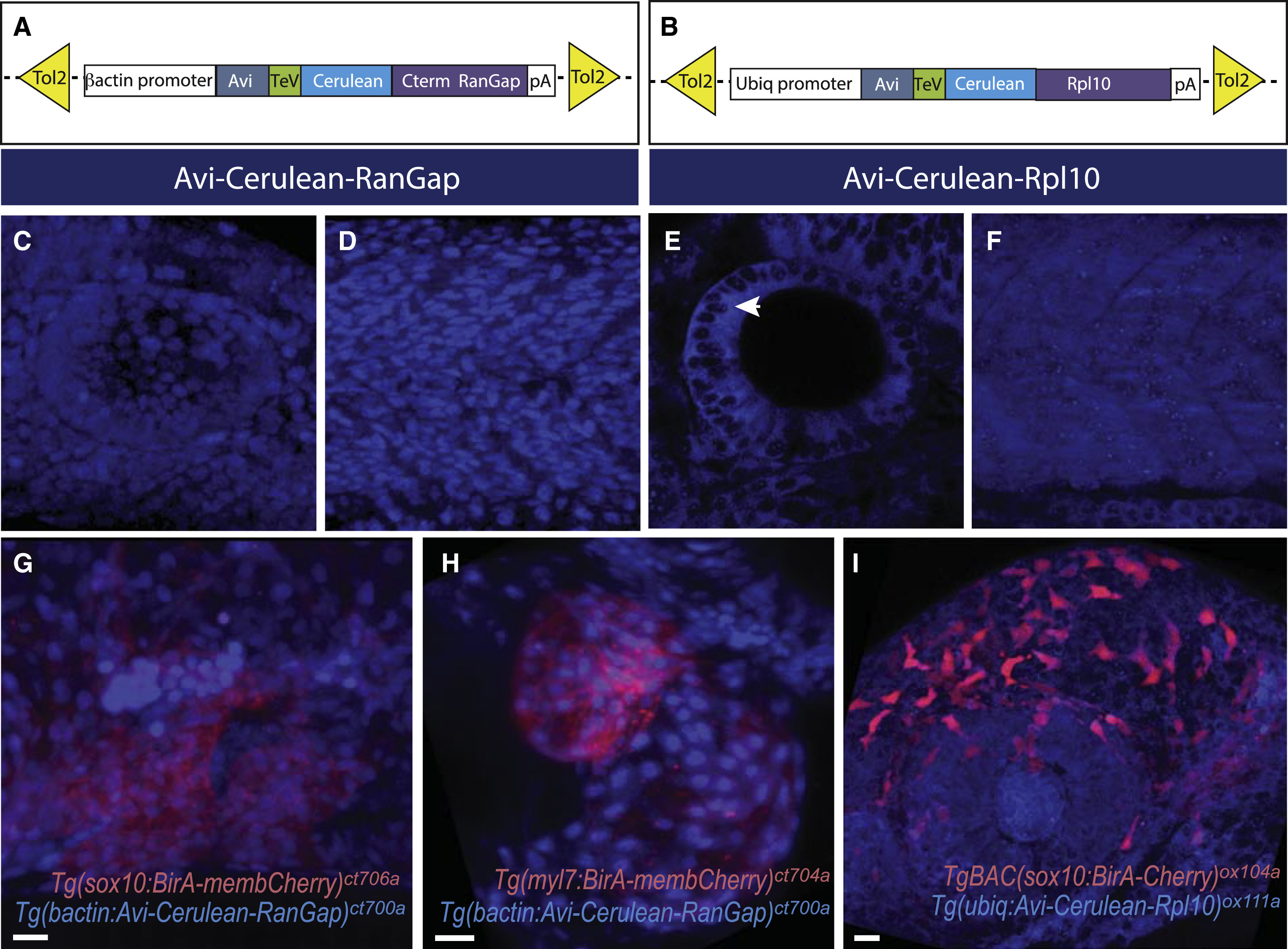
(A and B) Schematic of Avi-tagged constructs for generating nuclear effector (nucAvi) (A) and ribosome effector (riboAvi) (B).
(C–F) Confocal 3D projection of nucAvi (C and D) and riboAvi (E and F) expression in the developing inner ear (C and E) and somite (D and F) at 32 hpf. Arrow points to nucleoli.
(G–I) Confocal 3D projection of BirA driver (G and I in NC; H in myocardium, red) and Avi effector (G,H, nucAvi, and I, riboAvi, blue). Scale bars, 20μm.
Acknowledgments
This image is the copyrighted work of the attributed author or publisher, and
ZFIN has permission only to display this image to its users.
Additional permissions should be obtained from the applicable author or publisher of the image.
Full text @ Cell Rep.