
|
Fig. S5
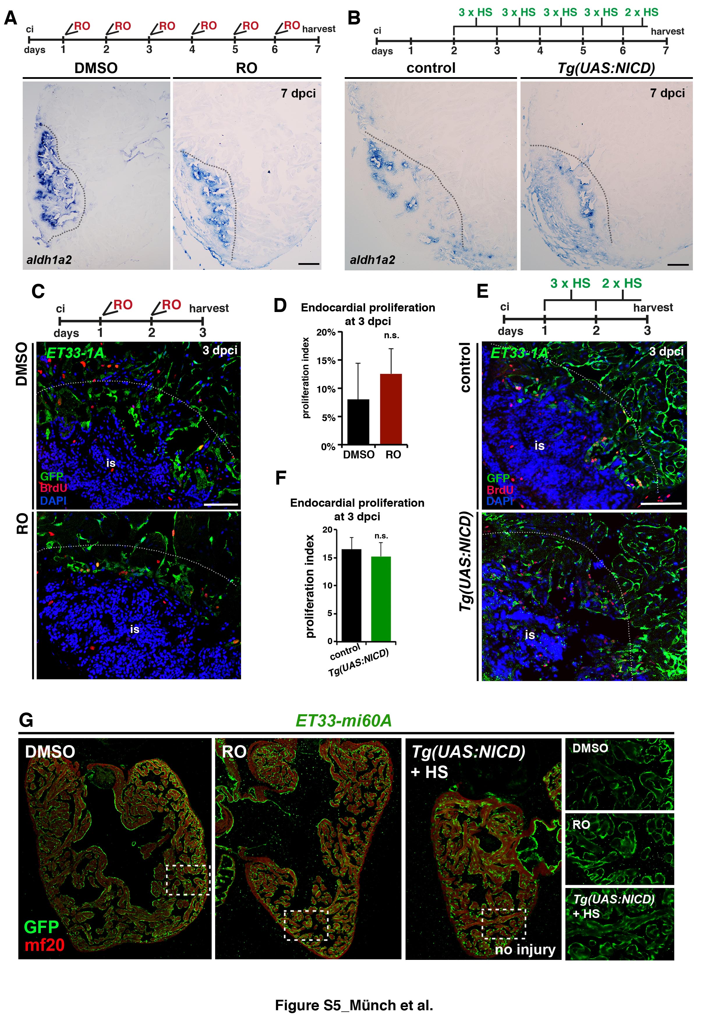
Notch signalling modulation does not affect aldh1a2 expression or proliferation (A,B) ISH for aldh1a2 on sections of hearts treated with DMSO or RO or from heat-shocked Tg(UAS:NICD) and control fish, showing no differences of expression. RO-treatment and heat shock regimes are indicated on top. (C) IHC against BrdU and GFP on heart sections of ET33-1a transgenic fish (3 dpci), showing BrdU incorporation by GFP+ endocardial cells adjacent to the injury site (is). (D) Quantification of BrdU+/ GFP+ cell ratio in hearts of fish treated with DMSO or RO, indicating no difference in endocardial cell proliferation (mean± s.d, t-test, not significant). (E) IHC against BrdU and GFP on heart sections of ET33-1a transgenic fish alone or crossed with Tg(UAS:NICD) at 3 dpci, showing BrdU incorporation by GFP+ endocardial cells adjacent to the injury site (is). (F) BrdU+/ GFP+ cell ratio in hearts of ET33-1a and Tg(UAS:NICD); ET33-1a transgenic fish, indicating no difference in endocardial cell proliferation (mean± s.d, t-test, not significant). (G) IHC for GFP and mf20 on sections of hearts treated with DMSO or RO or from heat-shocked Tg(UAS:NICD), showing no differences of expression. Boxed areas are magnified on the right. Hearts were treated for 3 days with RO, DMSO or heat shocks. Scale bars: (A) 100 μm in all panels.