
|
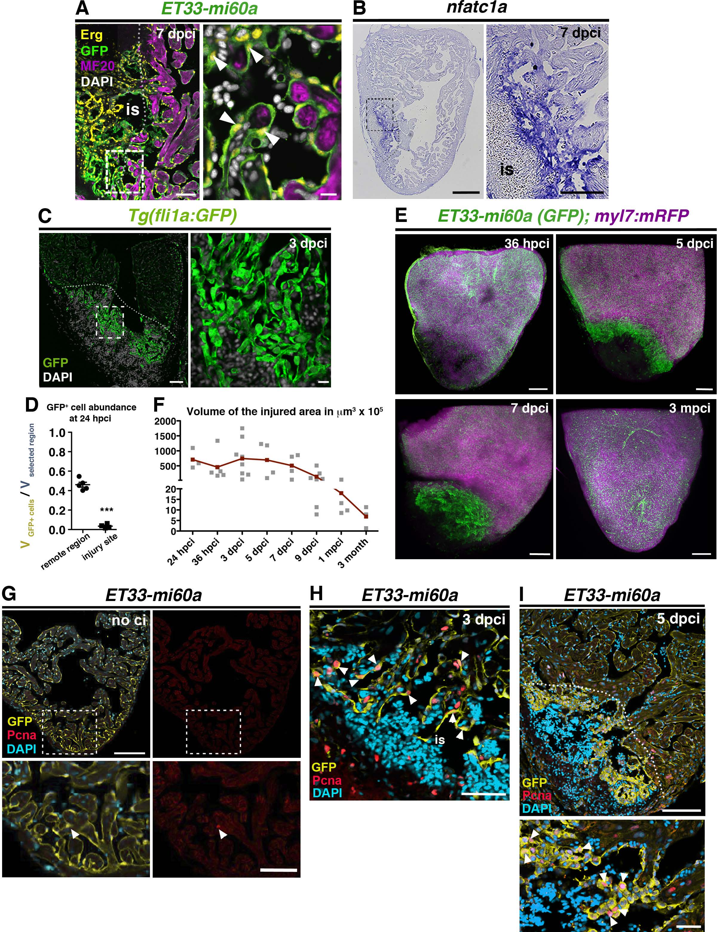
Fig. S1
Activated endocardium in the injury site (A) IHC against ERG, GFP and MF20 on sections of ET33-mi60a transgenic hearts (7 dpci), showing GFP+ endocardial/endothelial cells within and lining the injury site (is) (white arrowheads). The boxed area is magnified in the right-hand panel. (B) ISH against nfatc1a at 7 dpci, showing strong expression within the injury site (is). (C) IHC against GFP on sections of Tg(fli1a:GFP) transgenic hearts (3 dpci) showing endocardial cells at the injury site. The boxed area is magnified in the right-hand panel. (D) Scatter plot showing the relative volume occupied by GFP+ cells in a selected region of the remote region and the injury site at 24 hpci. Values of endocardial volume at the injury site are also represented in Figure 1L (black line = mean ± s.d, t-test, ***P<0.005). (E) Volume rendering of injured ventricles of ET33mi-60A; myl7mRFP hearts, with endocardium labelled green and myocardium magenta, at indicated time points. Volume rendering movies are available in the expanded view section. (F) Scatter plot showing the evolution of injury-site volume after cryoinjury in ET33-mi60a; myl7:mRFP transgenic hearts, quantified with IMARIS. (G-I) IHC against GFP and Pcna on sections of ET33-mi60a transgenic hearts (no ci, 3 dpci, 5dpci), showing almost any Pcna-expressing endocardial cell in the non-injured heart but high abundance of proliferating cells within the injury site (is) at 3 and 5 dpci (white arrowheads). The boxed area is magnified in the right hand panel. The dotted lines demarcate the injured tissue. Scale bars: 50 µm in A; 200 µm in B, E; 100 µm in C, G, H, I; 5 µm in magnified views in A, 100 µm in B, 20 µm in C,G, I.