
|
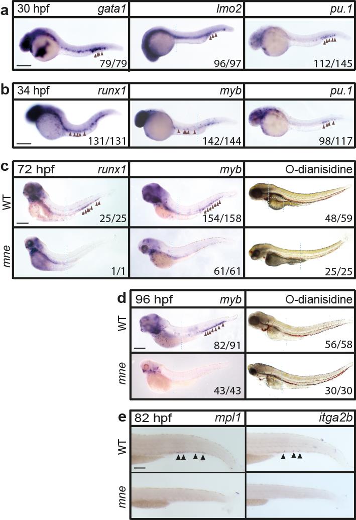
Fig. S2
Depletion of definitive haemopoiesis in mne (supports Fig. 1b)
Whole-mount in situ hybridization showing: a. Erythromyeloid specification marked by gata1, lmo2 and pu.1 expression proceeds in mne;
b. HSC specification marked by runx1 and myb proceeds in mne.
c,d. At 72 and 96 hpf, HSCs are depleted in mne while O-dianisidine staining shows haemoglobinised erythrocyte abundance is unaffected;
e. Decreased numbers of cells expressing mpl and itga2b shows that thrombocytes are depleted in mne. Where head and tail images of the same embryo were spliced to maintain in-focus focal plane, a dashed line indicates the junction; panels a-e scale bar = 200 μm.