
|
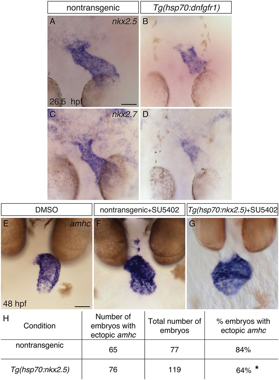
Fig. 9
FGF signaling functions upstream of Nkx genes to repress amhc expression in the ventricle. (A-D) In situ hybridization showing nkx2.5 (A,B) and nkx2.7 (C,D) expression in dorsal views at 26.5 hpf, following heat shock at 18 hpf. Nontransgenic embryos (A,C) exhibit robust expression of nkx2.5 (n=24) and nkx2.7 (n=12), and Tg(hsp70:dnfgfr1) embryos (B,D) exhibit reduced expression of nkx2.5 (n=24) and nkx2.7 (n=12). This reflects a reduced amount of ventricular tissue in Tg(hsp70:dnfgfr1) embryos (compare with Fig. 4M), as well as lower expression levels within this tissue. (E-G) In situ hybridization showing amhc expression at 48 hpf in nontransgenic (F) and Tg(hsp70:nkx2.5) (E,G) embryos treated with DMSO (E) or SU5402 (F,G); frontal views. In some cases (G), overexpression of nkx2.5 represses ectopic amhc expression in Tg(hsp70:nkx2.5) embryos that were treated with SU5402 at 18 hpf and subsequently heat shocked at 24 hpf. (H) Table reports the results of experiments gauging whether amhc expression in the ventricle of SU5402-treated embryos can be repressed by overexpression of nkx2.5. Asterisk indicates a statistically significant difference in the frequency of detecting ectopic amhc expression, compared to nontransgenic siblings (P=0.0019, Fisher’s exact test), representing partial rescue of the SU5402-treated phenotype. The degree of rescue was not enhanced when we used embryos carrying two copies of Tg(hsp70:nkx2.5) or when we administered two rounds of heat shock (data not shown). Scale bars: 50 μm.