
|
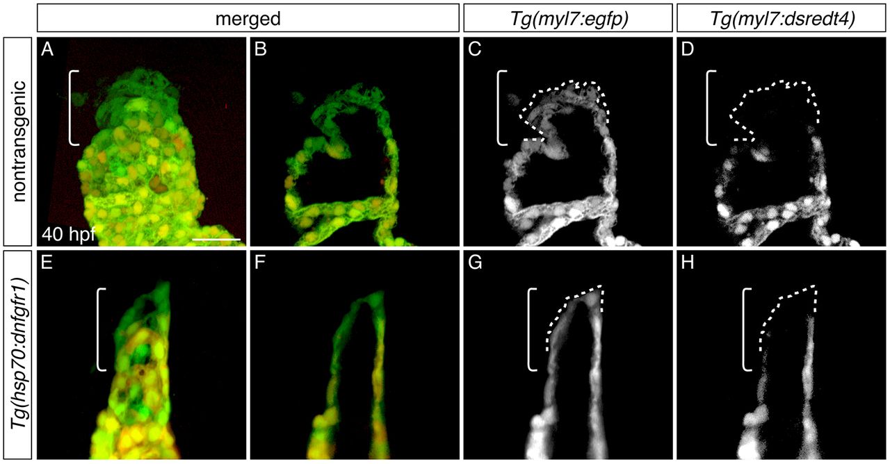
Fig. 7
Late-differentiating cardiomyocytes contribute to the heart after inhibition of FGF signaling. (A-H) Three-dimensional reconstructions (A,E) and single optical sections (B-D,F-H) of live embryos carrying Tg(myl7:egfp) and Tg(myl7:dsredt4); lateral views at 40 hpf, after heat shock at 18 hpf. Owing to the differential protein-folding kinetics of eGFP and dsRed (Lepilina et al., 2006), reporter transgene expression distinguishes early-differentiating (eGFP+dsRed+) and late-differentiating cardiomyocytes (eGFP+dsRed−) (de Pater et al., 2009). As in nontransgenic embryos (A-D, n=6), late-differentiating cardiomyocytes (eGFP+dsRed−, dashed outlines) contribute to the arterial pole (brackets) in Tg(hsp70:dnfgfr1) embryos (E-H; n=8). Scale bar: 30 μm.