
|
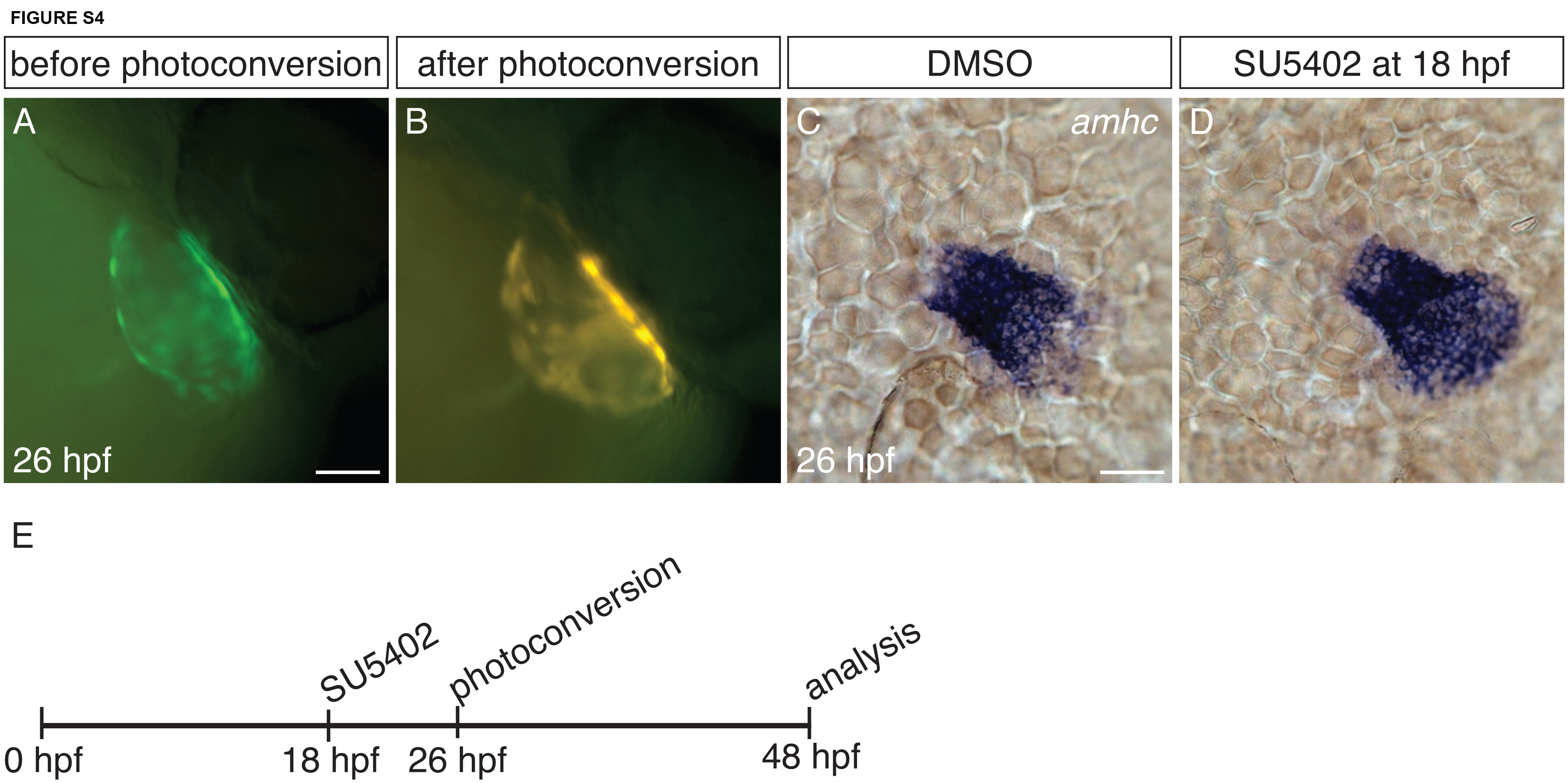
Fig. S4
Tg(amhc:dendra) facilitates tracking of amhc-expressing cells. (A-B) Tg(amhc:dendra) hearts before (A) and after (B) photoconversion at 26 hpf, lateral views. Tg(amhc:dendra) expression recapitulates the expression of the reporter transgene Tg(amhc:eGFP) (Zhang et al., 2013) and the expression of amhc itself (Fig. 4A-D): it is expressed throughout the atrium. Upon photoconversion, Dendra fluorescence is converted from green to red. Photoconversion at 26 hpf permits labeling of amhc-expressing cells throughout the atrium, and these photoconverted cells can then be detected at later stages (Fig. 6). (C-D) In situ hybridization depicts amhc expression in dorsal views of DMSO-treated and SU5402-treated embryos at 26 hpf. At 26 hpf, amhc expression in SU5402-treated embryos (D) is limited to the atrium; ectopic amhc-expressing cells are not yet visible at this stage.(E) Timeline of experimental design. Since amhc expression is not found in the ventricle of SU5402-treated embryos at 26 hpf, photoconversion at this stage permits examination of whether the ectopic amhc-expressing cells that appear later are derived from within the atrium or the AVC. Scale bars: 50 μm.