
Image
|
Figure Caption
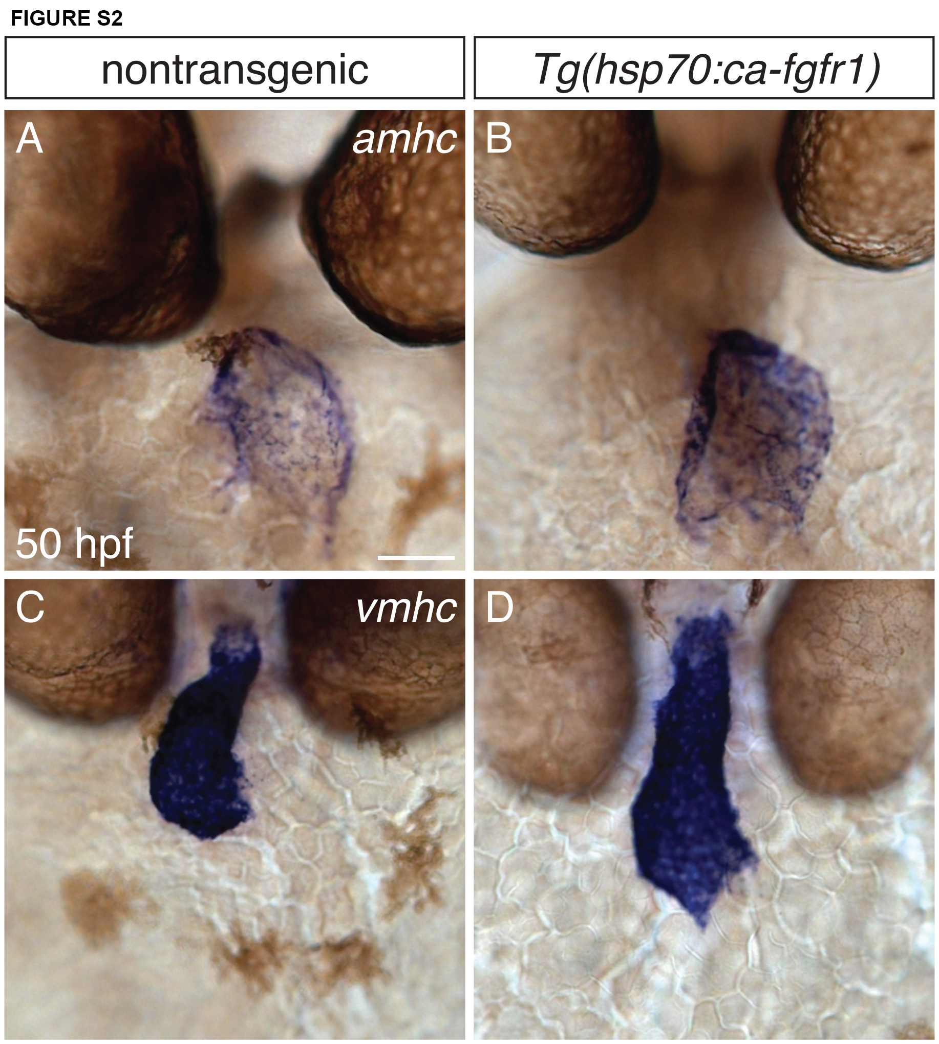
Fig. S2
Distribution of amhc and vmhc expression is unaffected by increased FGF signaling. (A-D) In situ hybridization depicts frontal views of amhc (A,B) and vmhc (C,D) expression at 50 hpf, following heat shock at 18 hpf. Nontransgenic embryos (A,C) show stereotypical expression patterns of amhc and vmhc (n=10). Increased FGF signaling in Tg(hsp70l:ca-fgfr1) embryos (B,D) results in slight morphological changes, consistent with prior observations (Marques et al., 2008). However, increased FGF signaling does not repress amhc in the atrium or result in ectopic vmhc expression (n=10). Scale bar: 50 μm.
Acknowledgments
This image is the copyrighted work of the attributed author or publisher, and
ZFIN has permission only to display this image to its users.
Additional permissions should be obtained from the applicable author or publisher of the image.
Full text @ Development