
|
Fig. S1
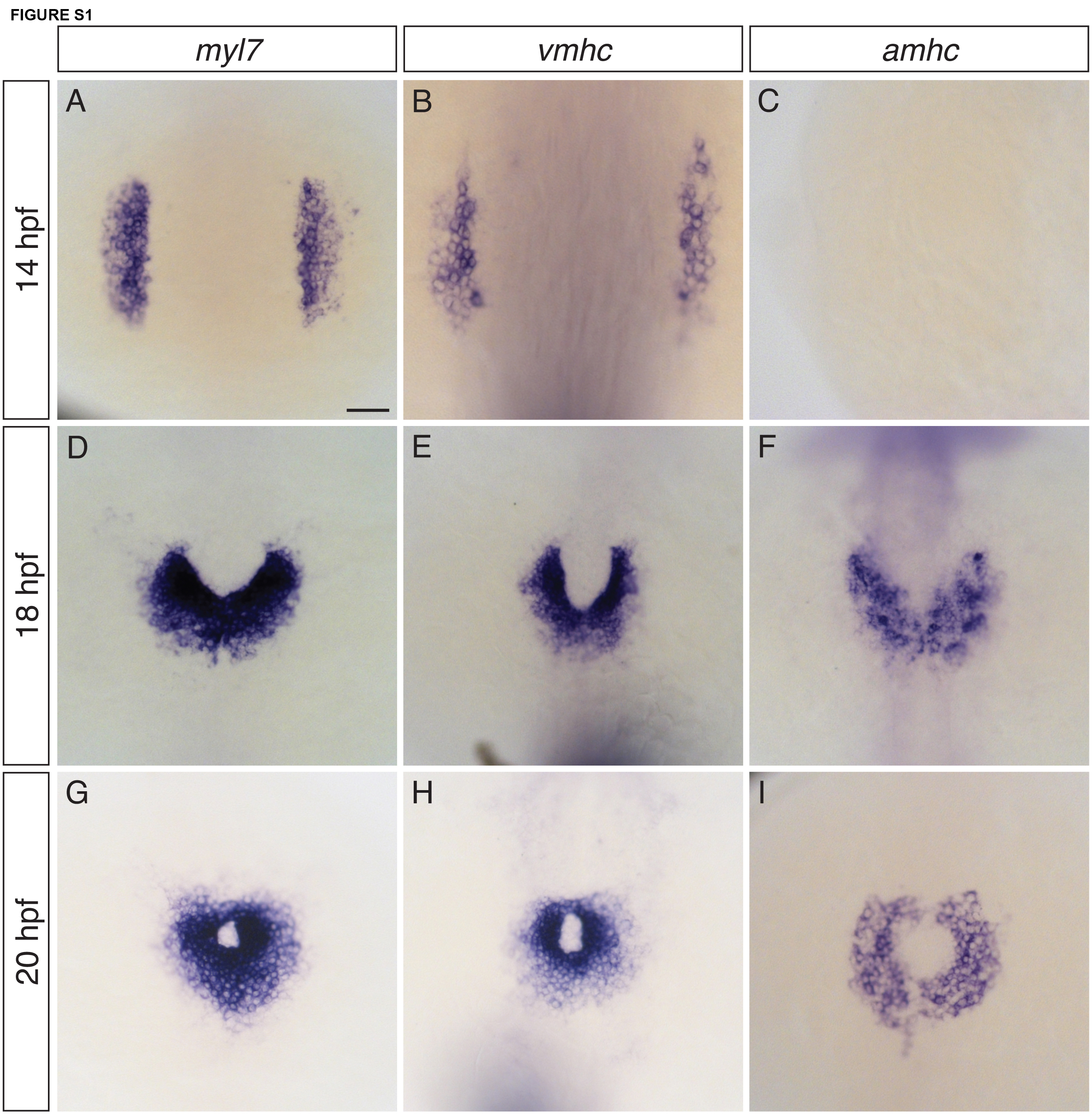
Expression of both vmhc and amhc is initiated by 18 hpf. (A-I) In situ hybridization depicts expression of myl7 (A,D,G), vmhc (B,E,H), and amhc (C,F,I) in dorsal views of wild-type embryos from 14-20 hpf (n=10 for each condition). (A,D,G) myl7 is expressed in all differentiated cardiomyocytes (Yelon et al., 1999). At 14 hpf, a subset of myl7-expressing cells express vmhc (B) and amhc is not yet expressed (C) (Yelon et al., 1999). By 18 hpf, cardiac fusion is underway, and vmhc (E) and amhc (F) are expressed in relatively medial and lateral populations of cardiomyocytes, corresponding to the locations of ventricular and atrial precursors (Schoenebeck et al., 2007). By 20 hpf, cardiomyocytes fuse at the midline to form a cardiac cone (Yelon et al., 1999), and vmhc (H) and amhc (I) expression are expressed in relatively central and peripheral populations of ventricular and atrial cardiomyocytes (Berdougo et al., 2003).
We do not consider either vmhc or amhc to be expressed in a strictly chamberspecific fashion: for example, low levels of vmhc are present in atrial cells at early stages (Berdougo et al., 2003; Yelon et al., 1999), and a subset of cells at the apex of the ventricle have been shown to express amhc (Foglia et al., 2016). Even so, the differences between the vmhc and amhc expression patterns point to the initiation of distinct differentiation programs in ventricular and atrial precursor populations, beginning as early as 18 hpf. Scale bar: 50 μm.