
|
Fig. 1
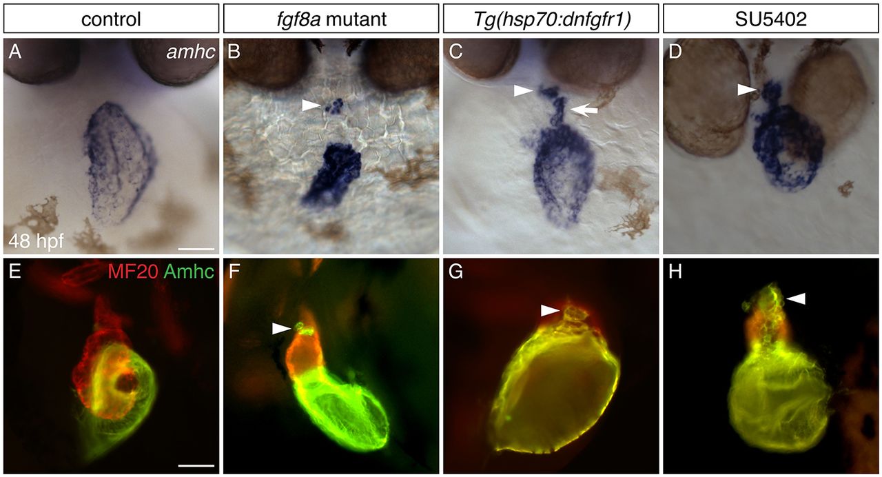
Inhibition of FGF signaling results in ectopic amhc expression in the ventricle. (A-D) In situ hybridization showing amhc expression in frontal views at 48 hpf. (A) Wild-type embryos express amhc in the atrium. Similar phenotypes are seen in all controls examined: wild-type siblings of fgf8a mutants, nontransgenic siblings following heat shock, and DMSO-treated siblings. (B) In fgf8a mutants, amhc expression is found not only in the atrium but also in a small cluster of ventricular cells (arrowhead; n=32/46 mutants). (C) More ectopic amhc-expressing cells are induced in Tg(hsp70:dnfgfr1) embryos following heat shock at 18 hpf (n=15/15); these cells are typically seen along the inner curvature (arrow) and at the arterial pole (arrowhead) of the ventricle. (D) Similarly, ectopic amhc-expressing cells can be induced by exposure to SU5402 starting at 18 hpf (arrowhead; n=15/15). The relatively mild phenotype of fgf8a mutants (B), compared with the more striking presence of ectopic cells in Tg(hsp70:dnfgfr1)-expressing and SU5402-treated embryos (C,D), suggests that additional FGF ligands might work together with fgf8a to prevent inappropriate amhc expression. (E-H) Immunofluorescence with MF20 (marks the myocardium; red) and S46 (recognizes Amhc; green) showing cardiac morphology and Amhc localization in lateral views at 48 hpf. In contrast to wild type (E), ectopic Amhc (arrowheads) is seen in fgf8a mutants (F), Tg(hsp70:dnfgfr1) embryos after heat shock at 18 hpf (G) and embryos treated with SU5402 from 18-30 hpf (H) (n>10 each). Scale bars: 50 μm.