
|
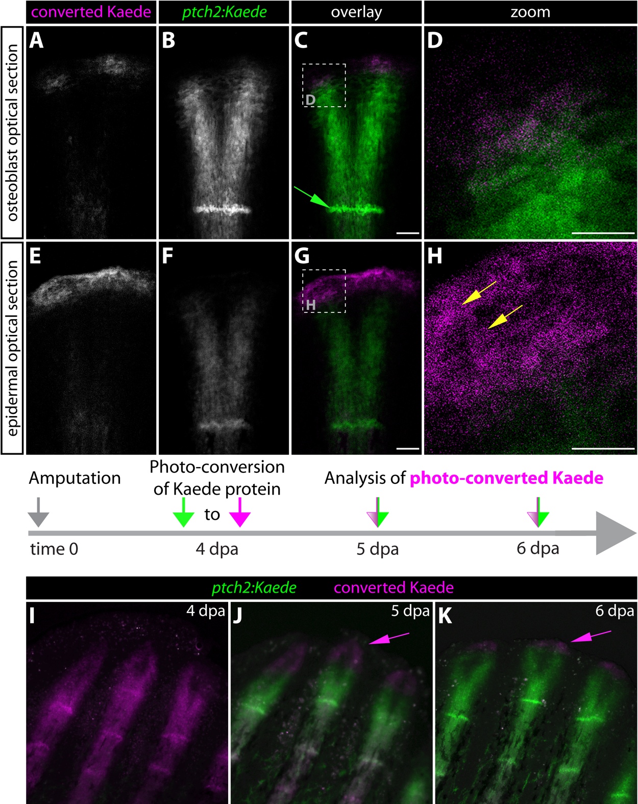
Fig. S2
The tip of regenerating fins contains previously Hedgehog/Smoothenedresponsive basal epidermal cells that subsequently moved further distally. (A-H) Kaede expression in the caudal fin of a 5 dpa ptch2:Kaede fish 24 hours after photoconversion. Images are selected single optical sections from a confocal z-stack of the same ray. (A-D) Optical section focused on the osteoblast layer. Dashed box in C indicates the region shown at higher magnification in panel D. Converted Kaede protein is exclusively found proximal to osteoblasts of the latest formed joint (green arrow). (E-H) Epidermal-focused optical section from the confocal z-stack. Dashed box in G indicates the zoomed region shown in H. (H) Yellow arrows point to converted Kaede protein at the distal tip of the regenerate. (I-K) Kaede expression at 5 and 6 dpa in ptch2:Kaede fish after photoconverting at 4 dpa. Previously Hh/Smo-responsive cells display converted Kaede in magenta, cells with both converted and nascent Kaede are white, and cells that transmitted Hh/Smo signals only after conversion show nascent Kaede alone in green. Magenta arrows indicate the extreme distal and previously Hh/Smo-responsive basal epidermal cells. Scale bars for D and H: 25 μm. All other scale bars: 50 μm.