
|
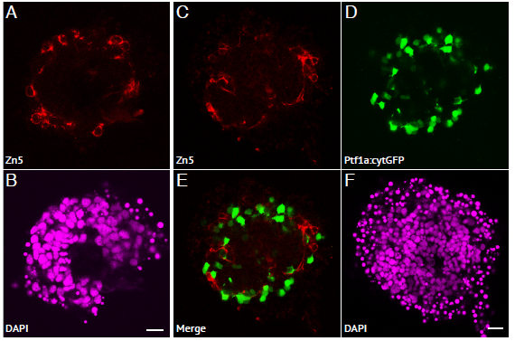
Fig. S4
RGC cells are positioned in the outer layer of zebrafish retinal aggregates.
(A-B) Aggregates cultured using the WT zebrafish strain, and stained with Zn5 primary antibody (marking RGCs) and DAPI. (A) RGCs can be seen positioned in the outer layer of the aggregate, and are extending axonal projections into the aggregate (arrows). (B) DAPI. Scale bar = 10μm. (C-F) Aggregates cultured using the Ptf1a:cytGFP zebrafish line, and stained with Zn5 primary antibody (marking RGCs) and DAPI. (C) RGCs can be seen positioned in the outer layer of the aggregate, and are extending axonal projections into the aggregate (arrows). (D) Ptf1a:cytGFP cells (namely ACs) are positioned in a ring around the outside of the aggregate. (E) Merge of (C and D) showing Zn5+ RGCs positioned in the outer layer, amongst the Ptf1a:cytGFP expressing cells. (F) DAPI. Scale bar = 10μm.