
|
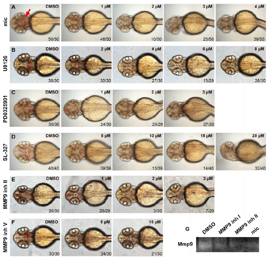
Fig. S7
Either MMP9 or pErk inhibitors decrease brain hemorrhage in fn40a mutants. Live imaging is shown for fn40a embryos treated by DMSO or small-molecule inhibitors. The hemorrhage rate of fn40a mutants was suppressed by miconazole (1 to 4 µmol/L (μM) mic, A), MEK inhibitor (2 to 8 µmol/L U0126, B; 1 to 3 µmol/L PD0325901, C; and 5 to 20 µmol/L SL-327, D), or MMP9 inhibitor II (1 to 3 µmol/L, E) and MMP9 inhibitor V (5 to 10 µmol/L, F) compared control embryos treated with DMSO. Embryos were orientated anterior on left with the dorsal view. Red arrow indicates the hemorrhage region. The concentrations of each compound were shown on upper right. The numbers on lower right show the phenotypical ones out of total scored embryos. (G) MMP9 zymography assay revealed that either MMP9 inhibitor I (3 µmol/L), MMP9 inhibitor II (3 µmol/L), or miconazole (3 µmol/L) decreased Mmp9 activities of homozygous fn40a mutants compared with DMSOtreated mutants. This represents one of two independent experiments.