
Image
|
Figure Caption
Fig. S9
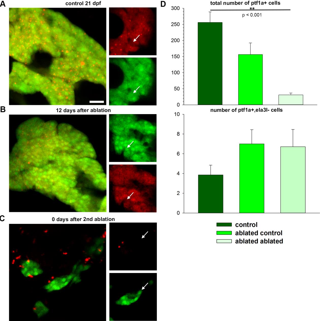
Ptf1a+ cells are maintained during regeneration. Confocal projections and single plane images (smaller images right to the projections) of A: untreated 21 dpf larva displaying mostly E2Crimson+ cells also positive for ptf1a:GFP; B: larva 12 days after an ablation of exocrine cells from 7 to 9dpf; C: larva at 0 dpa, treated a second time with Dim from 10 to 12 dpa showing ptf1a:GFP+ cells, not expressing E2Crimson; D: total number of ptf1a+ cells in control larvae (n=7), regenerating larvae (n=6) and double ablated larvae (n=7) and number of ptf1a+, ela3l- cells. Scale bar: 20μm.
Acknowledgments
This image is the copyrighted work of the attributed author or publisher, and
ZFIN has permission only to display this image to its users.
Additional permissions should be obtained from the applicable author or publisher of the image.
Full text @ Dis. Model. Mech.